6WLZ

The V1 region of human V-ATPase in state 1 (focused refinement)


Experimental Data Snapshot

  • Method: ELECTRON MICROSCOPY
  • Resolution: 2.90 Å
  • Aggregation State: PARTICLE 
  • Reconstruction Method: SINGLE PARTICLE 

wwPDB Validation   3D Report Full Report


This is version 1.3 of the entry. See complete history


Literature

Structures of a Complete Human V-ATPase Reveal Mechanisms of Its Assembly.

Wang, L.Wu, D.Robinson, C.V.Wu, H.Fu, T.M.

(2020) Mol Cell 80: 501

  • DOI: https://doi.org/10.1016/j.molcel.2020.09.029
  • Primary Citation of Related Structures:  
    6WLZ, 6WM4

  • PubMed Abstract: 

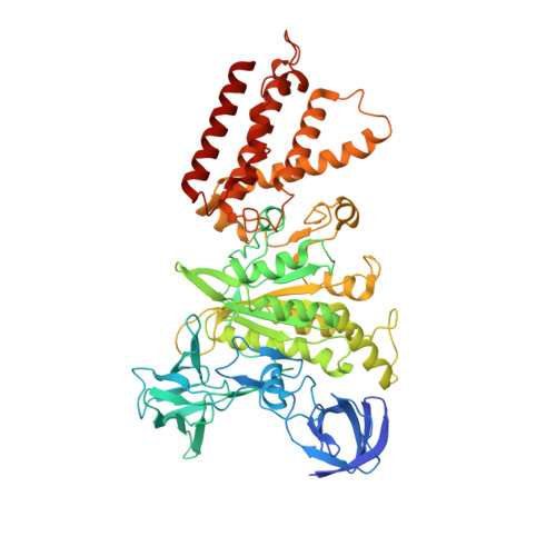
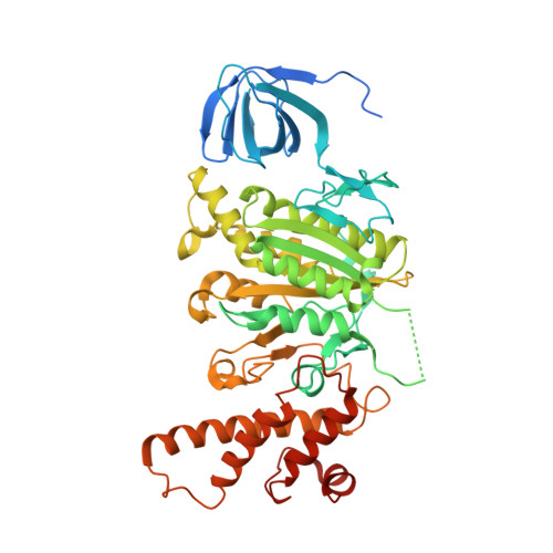
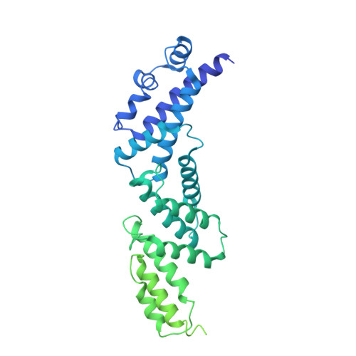
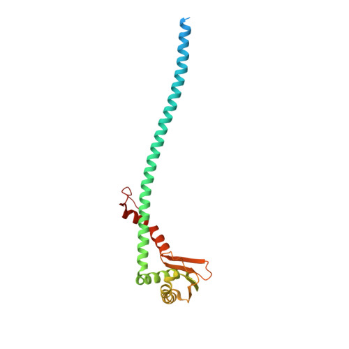
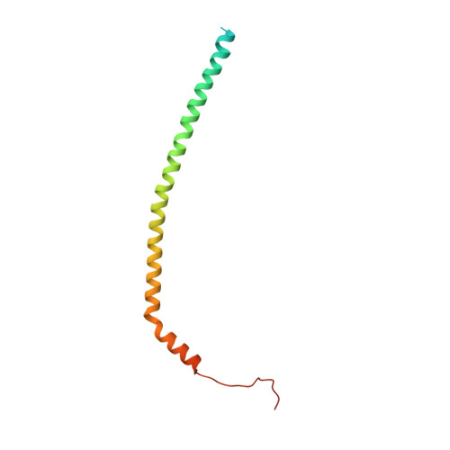
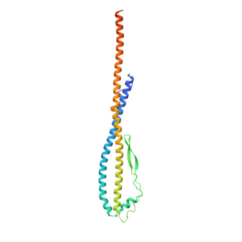
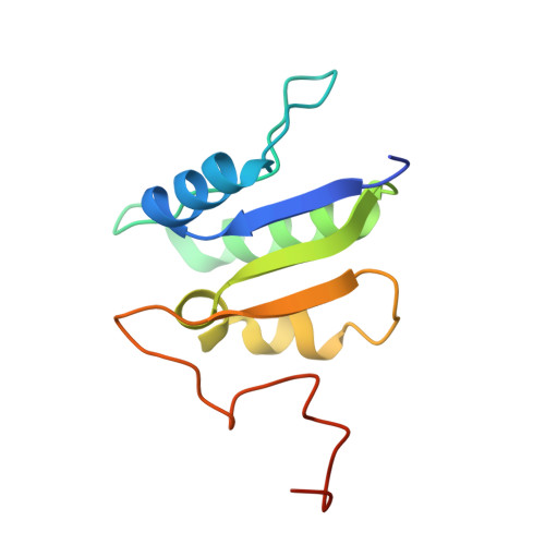
    Vesicular- or vacuolar-type adenosine triphosphatases (V-ATPases) are ATP-driven proton pumps comprised of a cytoplasmic V 1 complex for ATP hydrolysis and a membrane-embedded V o complex for proton transfer. They play important roles in acidification of intracellular vesicles, organelles, and the extracellular milieu in eukaryotes. Here, we report cryoelectron microscopy structures of human V-ATPase in three rotational states at up to 2.9-Å resolution. Aided by mass spectrometry, we build all known protein subunits with associated N-linked glycans and identify glycolipids and phospholipids in the V o complex. We define ATP6AP1 as a structural hub for V o complex assembly because it connects to multiple V o subunits and phospholipids in the c-ring. The glycolipids and the glycosylated V o subunits form a luminal glycan coat critical for V-ATPase folding, localization, and stability. This study identifies mechanisms of V-ATPase assembly and biogenesis that rely on the integrated roles of ATP6AP1, glycans, and lipids.


  • Organizational Affiliation

    Department of Biological Chemistry and Molecular Pharmacology, Harvard Medical School, Boston, MA 02115, USA; Program in Cellular and Molecular Medicine, Boston Children's Hospital, Boston, MA 02115, USA. Electronic address: wang@hkl.hms.harvard.edu.


Macromolecules
Find similar proteins by:  (by identity cutoff)  |  3D Structure
Entity ID: 1
MoleculeChains Sequence LengthOrganismDetailsImage
V-type proton ATPase catalytic subunit A
A, B, C
617Homo sapiensMutation(s): 0 
EC: 7.1.2.2
Membrane Entity: Yes 
UniProt & NIH Common Fund Data Resources
Find proteins for P38606 (Homo sapiens)
Explore P38606 
Go to UniProtKB:  P38606
PHAROS:  P38606
GTEx:  ENSG00000114573 
Entity Groups  
Sequence Clusters30% Identity50% Identity70% Identity90% Identity95% Identity100% Identity
UniProt GroupP38606
Sequence Annotations
Expand
  • Reference Sequence
Find similar proteins by:  (by identity cutoff)  |  3D Structure
Entity ID: 2
MoleculeChains Sequence LengthOrganismDetailsImage
V-type proton ATPase subunit B, brain isoform
D, E, F
511Homo sapiensMutation(s): 0 
Membrane Entity: Yes 
UniProt & NIH Common Fund Data Resources
Find proteins for P21281 (Homo sapiens)
Explore P21281 
Go to UniProtKB:  P21281
PHAROS:  P21281
GTEx:  ENSG00000147416 
Entity Groups  
Sequence Clusters30% Identity50% Identity70% Identity90% Identity95% Identity100% Identity
UniProt GroupP21281
Sequence Annotations
Expand
  • Reference Sequence
Find similar proteins by:  (by identity cutoff)  |  3D Structure
Entity ID: 3
MoleculeChains Sequence LengthOrganismDetailsImage
SidKG [auth X],
H [auth Y],
I [auth Z]
573Legionella pneumophila subsp. pneumophila str. Philadelphia 1Mutation(s): 0 
UniProt
Find proteins for Q5ZWW6 (Legionella pneumophila subsp. pneumophila (strain Philadelphia 1 / ATCC 33152 / DSM 7513))
Explore Q5ZWW6 
Go to UniProtKB:  Q5ZWW6
Entity Groups  
Sequence Clusters30% Identity50% Identity70% Identity90% Identity95% Identity100% Identity
UniProt GroupQ5ZWW6
Sequence Annotations
Expand
  • Reference Sequence
Find similar proteins by:  (by identity cutoff)  |  3D Structure
Entity ID: 4
MoleculeChains Sequence LengthOrganismDetailsImage
V-type proton ATPase subunit E 1J,
M [auth I],
O [auth H]
226Homo sapiensMutation(s): 0 
Membrane Entity: Yes 
UniProt & NIH Common Fund Data Resources
Find proteins for P36543 (Homo sapiens)
Explore P36543 
Go to UniProtKB:  P36543
PHAROS:  P36543
GTEx:  ENSG00000131100 
Entity Groups  
Sequence Clusters30% Identity50% Identity70% Identity90% Identity95% Identity100% Identity
UniProt GroupP36543
Sequence Annotations
Expand
  • Reference Sequence
Find similar proteins by:  (by identity cutoff)  |  3D Structure
Entity ID: 5
MoleculeChains Sequence LengthOrganismDetailsImage
V-type proton ATPase subunit G 1K [auth M],
N [auth L],
P [auth K]
118Homo sapiensMutation(s): 0 
Membrane Entity: Yes 
UniProt & NIH Common Fund Data Resources
Find proteins for O75348 (Homo sapiens)
Explore O75348 
Go to UniProtKB:  O75348
PHAROS:  O75348
GTEx:  ENSG00000136888 
Entity Groups  
Sequence Clusters30% Identity50% Identity70% Identity90% Identity95% Identity100% Identity
UniProt GroupO75348
Sequence Annotations
Expand
  • Reference Sequence
Find similar proteins by:  (by identity cutoff)  |  3D Structure
Entity ID: 6
MoleculeChains Sequence LengthOrganismDetailsImage
V-type proton ATPase subunit DL [auth G]247Homo sapiensMutation(s): 0 
Membrane Entity: Yes 
UniProt & NIH Common Fund Data Resources
Find proteins for Q9Y5K8 (Homo sapiens)
Explore Q9Y5K8 
Go to UniProtKB:  Q9Y5K8
PHAROS:  Q9Y5K8
GTEx:  ENSG00000100554 
Entity Groups  
Sequence Clusters30% Identity50% Identity70% Identity90% Identity95% Identity100% Identity
UniProt GroupQ9Y5K8
Sequence Annotations
Expand
  • Reference Sequence
Find similar proteins by:  (by identity cutoff)  |  3D Structure
Entity ID: 7
MoleculeChains Sequence LengthOrganismDetailsImage
V-type proton ATPase subunit FQ [auth N]119Homo sapiensMutation(s): 0 
Membrane Entity: Yes 
UniProt & NIH Common Fund Data Resources
Find proteins for Q16864 (Homo sapiens)
Explore Q16864 
Go to UniProtKB:  Q16864
PHAROS:  Q16864
GTEx:  ENSG00000128524 
Entity Groups  
Sequence Clusters30% Identity50% Identity70% Identity90% Identity95% Identity100% Identity
UniProt GroupQ16864
Sequence Annotations
Expand
  • Reference Sequence
Small Molecules
Ligands 1 Unique
IDChains Name / Formula / InChI Key2D Diagram3D Interactions
ADP
Query on ADP

Download Ideal Coordinates CCD File 
R [auth C]ADENOSINE-5'-DIPHOSPHATE
C10 H15 N5 O10 P2
XTWYTFMLZFPYCI-KQYNXXCUSA-N
Experimental Data & Validation

Experimental Data

  • Method: ELECTRON MICROSCOPY
  • Resolution: 2.90 Å
  • Aggregation State: PARTICLE 
  • Reconstruction Method: SINGLE PARTICLE 

Structure Validation

View Full Validation Report


Entry History 

Deposition Data

Revision History  (Full details and data files)

  • Version 1.0: 2020-11-11
    Type: Initial release
  • Version 1.1: 2020-11-18
    Changes: Database references
  • Version 1.2: 2021-03-31
    Changes: Database references, Derived calculations, Source and taxonomy, Structure summary
  • Version 1.3: 2024-03-06
    Changes: Data collection, Database references, Derived calculations