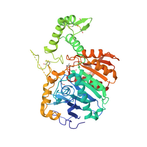
6WIA

CRYSTAL STRUCTURE OF HUMAN PROTECTIVE PROTEIN/CATHEPSIN A, DFP-INHIBITED (AGED)


Experimental Data Snapshot

  • Method: X-RAY DIFFRACTION
  • Resolution: 2.21 Å
  • R-Value Free: 0.224 
  • R-Value Work: 0.195 
  • R-Value Observed: 0.196 

Starting Model: experimental
View more details

wwPDB Validation   3D Report Full Report


This is version 2.1 of the entry. See complete history


Literature

Structural and kinetic evidence of aging after organophosphate inhibition of human Cathepsin A.

Bouknight, K.D.Jurkouich, K.M.Compton, J.R.Khavrutskii, I.V.Guelta, M.A.Harvey, S.P.Legler, P.M.

(2020) Biochem Pharmacol 177: 113980-113980

  • DOI: https://doi.org/10.1016/j.bcp.2020.113980
  • Primary Citation of Related Structures:  
    6WIA

  • PubMed Abstract: 

    Human Cathepsin A (CatA) is a lysosomal serine carboxypeptidase of the renin-angiotensin system (RAS) and is structurally similar to acetylcholinesterase (AChE). CatA can remove the C-terminal amino acids of endothelin I, angiotensin I, Substance P, oxytocin, and bradykinin, and can deamidate neurokinin A. Proteomic studies identified CatA and its homologue, SCPEP1, as potential targets of organophosphates (OP). CatA could be stably inhibited by low µM to high nM concentrations of racemic sarin (GB), soman (GD), cyclosarin (GF), VX, and VR within minutes to hours at pH 7. Cyclosarin was the most potent with a kinetically measured dissociation constant (K I ) of 2 µM followed by VR (K I  = 2.8 µM). Bimolecular rate constants for inhibition by cyclosarin and VR were 1.3 × 10 3 M -1 sec -1 and 1.2 × 10 3 M -1 sec -1 , respectively, and were approximately 3-orders of magnitude lower than those of human AChE indicating slower reactivity. Notably, both AChE and CatA bound diisopropylfluorophosphate (DFP) comparably and had K I DFP  = 13 µM and 11 µM, respectively. At low pH, greater than 85% of the enzyme spontaneously reactivated after OP inhibition, conditions under which OP-adducts of cholinesterases irreversibly age. At pH 6.5 CatA remained stably inhibited by GB and GF and <10% of the enzyme spontaneously reactivated after 200 h. A crystal structure of DFP-inhibited CatA was determined and contained an aged adduct. Similar to AChE, CatA appears to have a "backdoor" for product release. CatA has not been shown previously to age. These results may have implications for: OP-associated inflammation; cardiovascular effects; and the dysregulation of RAS enzymes by OP.


  • Organizational Affiliation

    Hampton University, 100 E Queen St, Hampton, VA 23668, United States.


Macromolecules
Find similar proteins by:  (by identity cutoff)  |  3D Structure
Entity ID: 1
MoleculeChains Sequence LengthOrganismDetailsImage
Lysosomal protective protein458Homo sapiensMutation(s): 0 
Gene Names: CTSAPPGB
EC: 3.4.16.5
UniProt & NIH Common Fund Data Resources
Find proteins for P10619 (Homo sapiens)
Explore P10619 
Go to UniProtKB:  P10619
PHAROS:  P10619
GTEx:  ENSG00000064601 
Entity Groups  
Sequence Clusters30% Identity50% Identity70% Identity90% Identity95% Identity100% Identity
UniProt GroupP10619
Glycosylation
Glycosylation Sites: 1Go to GlyGen: P10619-1
Sequence Annotations
Expand
  • Reference Sequence
Oligosaccharides

Help

Entity ID: 2
MoleculeChains Length2D Diagram Glycosylation3D Interactions
2-acetamido-2-deoxy-beta-D-glucopyranose-(1-4)-2-acetamido-2-deoxy-beta-D-glucopyranose
B, C
2N-Glycosylation
Glycosylation Resources
GlyTouCan:  G42666HT
GlyCosmos:  G42666HT
GlyGen:  G42666HT
Small Molecules
Ligands 3 Unique
IDChains Name / Formula / InChI Key2D Diagram3D Interactions
CD
Query on CD

Download Ideal Coordinates CCD File 
I [auth A]
J [auth A]
K [auth A]
L [auth A]
M [auth A]
I [auth A],
J [auth A],
K [auth A],
L [auth A],
M [auth A],
N [auth A]
CADMIUM ION
Cd
WLZRMCYVCSSEQC-UHFFFAOYSA-N
GOL
Query on GOL

Download Ideal Coordinates CCD File 
O [auth A]GLYCEROL
C3 H8 O3
PEDCQBHIVMGVHV-UHFFFAOYSA-N
ACT
Query on ACT

Download Ideal Coordinates CCD File 
D [auth A],
E [auth A],
F [auth A],
G [auth A],
H [auth A]
ACETATE ION
C2 H3 O2
QTBSBXVTEAMEQO-UHFFFAOYSA-M
Modified Residues  1 Unique
IDChains TypeFormula2D DiagramParent
MIS
Query on MIS
A
L-PEPTIDE LINKINGC6 H14 N O6 PSER
Experimental Data & Validation

Experimental Data

  • Method: X-RAY DIFFRACTION
  • Resolution: 2.21 Å
  • R-Value Free: 0.224 
  • R-Value Work: 0.195 
  • R-Value Observed: 0.196 
  • Space Group: C 1 2 1
Unit Cell:
Length ( Å )Angle ( ˚ )
a = 91.273α = 90
b = 101.999β = 101.61
c = 48.114γ = 90
Software Package:
Software NamePurpose
REFMACrefinement
PDB_EXTRACTdata extraction
APEXdata reduction
APEXdata scaling
PHASERphasing

Structure Validation

View Full Validation Report


Entry History & Funding Information

Deposition Data


Funding OrganizationLocationGrant Number
Department of Defense (DOD, United States)United States#GW150096

Revision History  (Full details and data files)

  • Version 1.0: 2020-04-29
    Type: Initial release
  • Version 1.1: 2020-05-20
    Changes: Database references
  • Version 2.0: 2020-07-29
    Type: Remediation
    Reason: Carbohydrate remediation
    Changes: Advisory, Atomic model, Data collection, Derived calculations, Structure summary
  • Version 2.1: 2023-10-18
    Changes: Data collection, Database references, Refinement description, Structure summary