6WE3

Human PARP14 (ARTD8), catalytic fragment in complex with compound 3


Experimental Data Snapshot

  • Method: X-RAY DIFFRACTION
  • Resolution: 1.95 Å
  • R-Value Free: 0.273 
  • R-Value Work: 0.237 
  • R-Value Observed: 0.239 

wwPDB Validation   3D Report Full Report


Ligand Structure Quality Assessment 


This is version 1.2 of the entry. See complete history


Literature

A potent and selective PARP14 inhibitor decreases protumor macrophage gene expression and elicits inflammatory responses in tumor explants.

Schenkel, L.B.Molina, J.R.Swinger, K.K.Abo, R.Blackwell, D.J.Lu, A.Z.Cheung, A.E.Church, W.D.Kunii, K.Kuplast-Barr, K.G.Majer, C.R.Minissale, E.Mo, J.R.Niepel, M.Reik, C.Ren, Y.Vasbinder, M.M.Wigle, T.J.Richon, V.M.Keilhack, H.Kuntz, K.W.

(2021) Cell Chem Biol 28: 1158

  • DOI: https://doi.org/10.1016/j.chembiol.2021.02.010
  • Primary Citation of Related Structures:  
    6WE2, 6WE3, 6WE4

  • PubMed Abstract: 

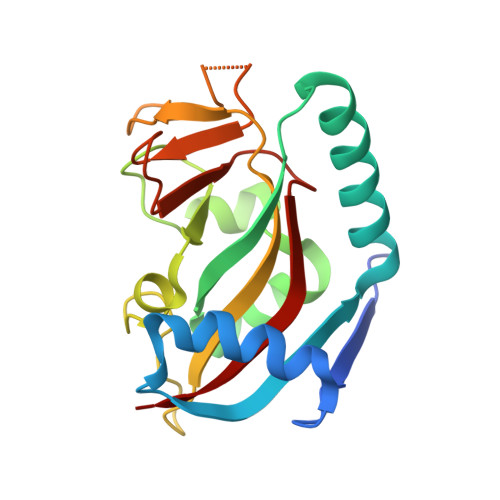
    PARP14 has been implicated by genetic knockout studies to promote protumor macrophage polarization and suppress the antitumor inflammatory response due to its role in modulating interleukin-4 (IL-4) and interferon-γ signaling pathways. Here, we describe structure-based design efforts leading to the discovery of a potent and highly selective PARP14 chemical probe. RBN012759 inhibits PARP14 with a biochemical half-maximal inhibitory concentration of 0.003 μM, exhibits >300-fold selectivity over all PARP family members, and its profile enables further study of PARP14 biology and disease association both in vitro and in vivo. Inhibition of PARP14 with RBN012759 reverses IL-4-driven protumor gene expression in macrophages and induces an inflammatory mRNA signature similar to that induced by immune checkpoint inhibitor therapy in primary human tumor explants. These data support an immune suppressive role of PARP14 in tumors and suggest potential utility of PARP14 inhibitors in the treatment of cancer.


  • Organizational Affiliation

    Department of Molecular Discovery, Ribon Therapeutics, Inc., Cambridge, MA 02140, USA; MOMA Therapeutics, Cambridge, MA 02142, USA.


Macromolecules
Find similar proteins by:  (by identity cutoff)  |  3D Structure
Entity ID: 1
MoleculeChains Sequence LengthOrganismDetailsImage
Protein mono-ADP-ribosyltransferase PARP14
A, B
194Homo sapiensMutation(s): 0 
Gene Names: PARP14BAL2KIAA1268
EC: 2.4.2
UniProt & NIH Common Fund Data Resources
Find proteins for Q460N5 (Homo sapiens)
Explore Q460N5 
Go to UniProtKB:  Q460N5
PHAROS:  Q460N5
GTEx:  ENSG00000173193 
Entity Groups  
Sequence Clusters30% Identity50% Identity70% Identity90% Identity95% Identity100% Identity
UniProt GroupQ460N5
Sequence Annotations
Expand
  • Reference Sequence
Small Molecules
Ligands 6 Unique
IDChains Name / Formula / InChI Key2D Diagram3D Interactions
TZ7 (Subject of Investigation/LOI)
Query on TZ7

Download Ideal Coordinates CCD File 
C [auth A],
P [auth B]
2-{[(trans-4-hydroxycyclohexyl)sulfanyl]methyl}-8-methylquinazolin-4(3H)-one
C16 H20 N2 O2 S
OSFGFBLRIXHPSO-HAQNSBGRSA-N
PGE
Query on PGE

Download Ideal Coordinates CCD File 
U [auth B]TRIETHYLENE GLYCOL
C6 H14 O4
ZIBGPFATKBEMQZ-UHFFFAOYSA-N
PEG
Query on PEG

Download Ideal Coordinates CCD File 
J [auth A],
V [auth B]
DI(HYDROXYETHYL)ETHER
C4 H10 O3
MTHSVFCYNBDYFN-UHFFFAOYSA-N
SO4
Query on SO4

Download Ideal Coordinates CCD File 
M [auth A],
N [auth A],
O [auth A]
SULFATE ION
O4 S
QAOWNCQODCNURD-UHFFFAOYSA-L
GOL
Query on GOL

Download Ideal Coordinates CCD File 
K [auth A],
L [auth A],
W [auth B],
X [auth B]
GLYCEROL
C3 H8 O3
PEDCQBHIVMGVHV-UHFFFAOYSA-N
EDO
Query on EDO

Download Ideal Coordinates CCD File 
D [auth A]
E [auth A]
F [auth A]
G [auth A]
H [auth A]
D [auth A],
E [auth A],
F [auth A],
G [auth A],
H [auth A],
I [auth A],
Q [auth B],
R [auth B],
S [auth B],
T [auth B]
1,2-ETHANEDIOL
C2 H6 O2
LYCAIKOWRPUZTN-UHFFFAOYSA-N
Experimental Data & Validation

Experimental Data

  • Method: X-RAY DIFFRACTION
  • Resolution: 1.95 Å
  • R-Value Free: 0.273 
  • R-Value Work: 0.237 
  • R-Value Observed: 0.239 
  • Space Group: C 2 2 21
Unit Cell:
Length ( Å )Angle ( ˚ )
a = 82.353α = 90
b = 144.656β = 90
c = 82.541γ = 90
Software Package:
Software NamePurpose
HKL-2000data scaling
REFMACrefinement
PDB_EXTRACTdata extraction
HKL-3000data reduction
REFMACphasing

Structure Validation

View Full Validation Report


Ligand Structure Quality Assessment 


Entry History 

Deposition Data

Revision History  (Full details and data files)

  • Version 1.0: 2021-03-24
    Type: Initial release
  • Version 1.1: 2021-09-01
    Changes: Database references
  • Version 1.2: 2023-10-18
    Changes: Data collection, Refinement description