6WA8

Crystal structure of the E. coli transcription termination factor Rho


Experimental Data Snapshot

  • Method: X-RAY DIFFRACTION
  • Resolution: 3.30 Å
  • R-Value Free: 0.296 
  • R-Value Work: 0.252 
  • R-Value Observed: 0.254 

wwPDB Validation   3D Report Full Report


Ligand Structure Quality Assessment 


This is version 1.1 of the entry. See complete history


Literature

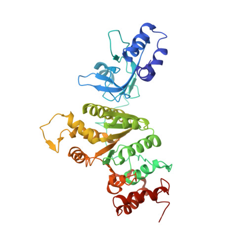
Crystal structure of the Escherichia coli transcription termination factor Rho.

Fan, C.Rees, D.C.

(2020) Acta Crystallogr F Struct Biol Commun 76: 398-405

  • DOI: https://doi.org/10.1107/S2053230X20010572
  • Primary Citation of Related Structures:  
    6WA8

  • PubMed Abstract: 

    During the crystal structure analysis of an ATP-binding cassette (ABC) transporter overexpressed in Escherichia coli, a contaminant protein was crystallized. The identity of the contaminant was revealed by mass spectrometry to be the Escherichia coli transcription terminator factor Rho, structures of which had been previously determined in different conformational states. Although Rho was present at only ∼1% of the target protein (a bacterial homolog of the eukaryotic ABC transporter of mitochondria from Novosphingobium aromaticivorans; NaAtm1), it preferentially crystallized in space group C2 as thin plates that diffracted to 3.30 Å resolution. The structure of Rho in this crystal form exhibits a hexameric open-ring staircase conformation with bound ATP; this characteristic structure was also observed on electron-microscopy grids of the NaAtm1 preparation.


  • Organizational Affiliation

    Division of Chemistry and Chemical Engineering, California Institute of Technology, 1200 East California Boulevard, Pasadena, CA 91125, USA.


Macromolecules
Find similar proteins by:  (by identity cutoff)  |  3D Structure
Entity ID: 1
MoleculeChains Sequence LengthOrganismDetailsImage
Transcription termination factor Rho
A, B, C, D, E
A, B, C, D, E, F
419Escherichia coli K-12Mutation(s): 0 
Gene Names: rhonitApsuArnsCsbaAtsub3783JW3756
EC: 3.6.4
UniProt
Find proteins for P0AG30 (Escherichia coli (strain K12))
Explore P0AG30 
Go to UniProtKB:  P0AG30
Entity Groups  
Sequence Clusters30% Identity50% Identity70% Identity90% Identity95% Identity100% Identity
UniProt GroupP0AG30
Sequence Annotations
Expand
  • Reference Sequence
Experimental Data & Validation

Experimental Data

  • Method: X-RAY DIFFRACTION
  • Resolution: 3.30 Å
  • R-Value Free: 0.296 
  • R-Value Work: 0.252 
  • R-Value Observed: 0.254 
  • Space Group: C 1 2 1
Unit Cell:
Length ( Å )Angle ( ˚ )
a = 161.745α = 90
b = 101.902β = 107.77
c = 184.016γ = 90
Software Package:
Software NamePurpose
PDB_EXTRACTdata extraction
PHENIXrefinement
PHASERphasing
Aimlessdata scaling
XDSdata reduction
JBluIce-EPICSdata collection

Structure Validation

View Full Validation Report


Ligand Structure Quality Assessment 


Entry History & Funding Information

Deposition Data


Funding OrganizationLocationGrant Number
Howard Hughes Medical Institute (HHMI)United States--

Revision History  (Full details and data files)

  • Version 1.0: 2020-09-16
    Type: Initial release
  • Version 1.1: 2023-10-18
    Changes: Data collection, Database references, Refinement description