


Plasmodium vivax and human hexokinases share similar active sites but display distinct quaternary architectures
Srivastava, S.S., Darling, J.E., Suryadi, J., Morris, J.C., Drew, M.E., Subramaniam, S.(2020) IUCrJ 7: 453-461
Experimental Data Snapshot
wwPDB Validation 3D Report Full Report

(2020) IUCrJ 7: 453-461
Entity ID: 1 | |||||
|---|---|---|---|---|---|
| Molecule | Chains | Sequence Length | Organism | Details | Image |
| Phosphotransferase | 505 | Plasmodium vivax | Mutation(s): 0 Gene Names: PVC01_110030900, PVP01_1125500, PVT01_110029900 EC: 2.7.1 | 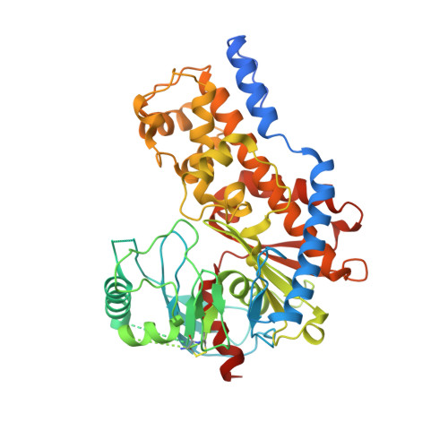 | |
UniProt | |||||
Find proteins for A5K274 (Plasmodium vivax (strain Salvador I)) Explore A5K274 Go to UniProtKB: A5K274 | |||||
Entity Groups | |||||
| Sequence Clusters | 30% Identity50% Identity70% Identity90% Identity95% Identity100% Identity | ||||
| UniProt Group | A5K274 | ||||
Sequence AnnotationsExpand | |||||
| |||||
| Task | Software Package | Version |
|---|---|---|
| MODEL REFINEMENT | PHENIX | |
| RECONSTRUCTION | RELION | 3.0 |
| Funding Organization | Location | Grant Number |
|---|---|---|
| Canada Excellence Research Chair Award | Canada | -- |


