


HIV Integrase core domain in complex with inhibitor (5-methyl-1-benzofuran-3-yl)acetic acid
Gorman, M.A., Parker, M.W.To be published.
Experimental Data Snapshot
Entity ID: 1 | |||||
|---|---|---|---|---|---|
| Molecule | Chains | Sequence Length | Organism | Details | Image |
| Integrase | 163 | Human immunodeficiency virus 1 | Mutation(s): 4 Gene Names: pol EC: 2.7.7 | 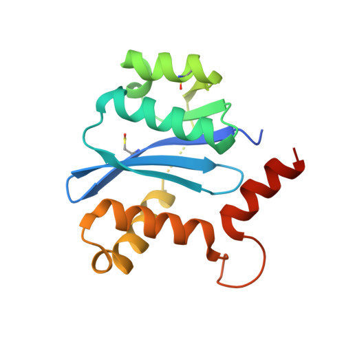 | |
UniProt | |||||
Entity Groups | |||||
| Sequence Clusters | 30% Identity50% Identity70% Identity90% Identity95% Identity100% Identity | ||||
| UniProt Group | P12497 | ||||
Sequence AnnotationsExpand | |||||
Reference Sequence | |||||
| Ligands 3 Unique | |||||
|---|---|---|---|---|---|
| ID | Chains | Name / Formula / InChI Key | 2D Diagram | 3D Interactions | |
| RQG (Subject of Investigation/LOI) Download:Ideal Coordinates CCD File | I [auth A] | (5-methyl-1-benzofuran-3-yl)acetic acid C11 H10 O3 DUMCTHVOZLFMDK-UHFFFAOYSA-N |  | ||
| IOD Download:Ideal Coordinates CCD File | B [auth A] C [auth A] D [auth A] E [auth A] F [auth A] | IODIDE ION I XMBWDFGMSWQBCA-UHFFFAOYSA-M |  | ||
| SO4 Download:Ideal Coordinates CCD File | J [auth A], K [auth A] | SULFATE ION O4 S QAOWNCQODCNURD-UHFFFAOYSA-L |  | ||
| Modified Residues 2 Unique | |||||
|---|---|---|---|---|---|
| ID | Chains | Type | Formula | 2D Diagram | Parent |
| CSD Query on CSD | A | L-PEPTIDE LINKING | C3 H7 N O4 S |  | CYS |
| CSO Query on CSO | A | L-PEPTIDE LINKING | C3 H7 N O3 S |  | CYS |
| Length ( Å ) | Angle ( ˚ ) |
|---|---|
| a = 46.288 | α = 90 |
| b = 46.288 | β = 90 |
| c = 140.096 | γ = 90 |
| Software Name | Purpose |
|---|---|
| REFMAC | refinement |
| Aimless | data reduction |
| Aimless | data scaling |
| REFMAC | phasing |


