6VP4 | pdb_00006vp4

Ethylene forming enzyme (EFE) in complex with Fe(II), L-arginine, and 2OG


Experimental Data Snapshot

  • Method: X-RAY DIFFRACTION
  • Resolution: 1.83 Å
  • R-Value Free: 
    0.197 (Depositor), 0.197 (DCC) 
  • R-Value Work: 
    0.174 (Depositor), 0.175 (DCC) 
  • R-Value Observed: 
    0.175 (Depositor) 

Starting Model: experimental
View more details

wwPDB Validation 3D Report Full Report

Validation slider image for 6VP4

Ligand Structure Quality Assessment 


This is version 1.3 of the entry. See complete history

Literature

An Iron(IV)-Oxo Intermediate Initiating l-Arginine Oxidation but Not Ethylene Production by the 2-Oxoglutarate-Dependent Oxygenase, Ethylene-Forming Enzyme.

Copeland, R.A.Davis, K.M.Shoda, T.K.C.Blaesi, E.J.Boal, A.K.Krebs, C.Bollinger Jr., J.M.

(2021) J Am Chem Soc 143: 2293-2303

  • DOI: https://doi.org/10.1021/jacs.0c10923
  • Primary Citation Related Structures: 
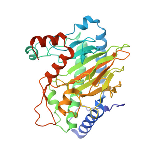
    6VP4, 6VP5

  • PubMed Abstract: 

    Ethylene-forming enzyme (EFE) is an ambifunctional iron(II)- and 2-oxoglutarate-dependent (Fe/2OG) oxygenase. In its major (EF) reaction, it converts carbons 1, 2, and 5 of 2OG to CO 2 and carbons 3 and 4 to ethylene, a four-electron oxidation drastically different from the simpler decarboxylation of 2OG to succinate mediated by all other Fe/2OG enzymes. EFE also catalyzes a minor reaction, in which the normal decarboxylation is coupled to oxidation of l-arginine (a required activator for the EF pathway), resulting in its conversion to l-glutamate semialdehyde and guanidine. Here we show that, consistent with precedent, the l-Arg-oxidation (RO) pathway proceeds via an iron(IV)-oxo (ferryl) intermediate. Use of 5,5-[ 2 H 2 ]-l-Arg slows decay of the ferryl complex by >16-fold, implying that RO is initiated by hydrogen-atom transfer (HAT) from C5. That this large substrate deuterium kinetic isotope effect has no impact on the EF:RO partition ratio implies that the same ferryl intermediate cannot be on the EF pathway; the pathways must diverge earlier. Consistent with this conclusion, the variant enzyme bearing the Asp191Glu ligand substitution accumulates ∼4 times as much of the ferryl complex as the wild-type enzyme and exhibits a ∼40-fold diminished EF:RO partition ratio. The selective detriment of this nearly conservative substitution to the EF pathway implies that it has unusually stringent stereoelectronic requirements. An active-site, like-charge guanidinium pair, which involves the l-Arg substrate/activator and is unique to EFE among four crystallographically characterized l-Arg-modifying Fe/2OG oxygenases, may serve to selectively stabilize the transition state leading to the unique EF branch.

Macromolecule Content 

  • Total Structure Weight: 159.76 kDa 
  • Atom Count: 11,760 
  • Modeled Residue Count: 1,343 
  • Deposited Residue Count: 1,400 
  • Unique protein chains: 1

Macromolecules

Find similar proteins by:|  3D Structure
Entity ID: 1
MoleculeChains  Sequence LengthOrganismDetailsImage
2-oxoglutarate-dependent ethylene/succinate-forming enzyme
A, B, C, D
350Pseudomonas savastanoi pv. phaseolicolaMutation(s): 0 
Gene Names: efe
EC: 1.13.12.19 (PDB Primary Data), 1.14.20.7 (PDB Primary Data)
UniProt
Find proteins for P32021 (Pseudomonas savastanoi pv. phaseolicola)
Explore P32021 
Go to UniProtKB:  P32021
Entity Groups
Sequence Clusters30% Identity50% Identity70% Identity90% Identity95% Identity100% Identity
UniProt GroupP32021
Sequence Annotations
Expand
Reference Sequence

Small Molecules

Ligands 4 Unique
IDChains Name / Formula / InChI Key2D Diagram3D Interactions
B3P

Query on B3P


Download:Ideal Coordinates CCD File
Q [auth D]2-[3-(2-HYDROXY-1,1-DIHYDROXYMETHYL-ETHYLAMINO)-PROPYLAMINO]-2-HYDROXYMETHYL-PROPANE-1,3-DIOL
C11 H26 N2 O6
HHKZCCWKTZRCCL-UHFFFAOYSA-N
ARG

Query on ARG


Download:Ideal Coordinates CCD File
G [auth A],
J [auth B],
M [auth C],
P [auth D]
ARGININE
C6 H15 N4 O2
ODKSFYDXXFIFQN-BYPYZUCNSA-O
AKG

Query on AKG


Download:Ideal Coordinates CCD File
F [auth A],
I [auth B],
L [auth C],
O [auth D]
2-OXOGLUTARIC ACID
C5 H6 O5
KPGXRSRHYNQIFN-UHFFFAOYSA-N
FE

Query on FE


Download:Ideal Coordinates CCD File
E [auth A],
H [auth B],
K [auth C],
N [auth D]
FE (III) ION
Fe
VTLYFUHAOXGGBS-UHFFFAOYSA-N

Experimental Data & Validation

Experimental Data

  • Method: X-RAY DIFFRACTION
  • Resolution: 1.83 Å
  • R-Value Free:  0.197 (Depositor), 0.197 (DCC) 
  • R-Value Work:  0.174 (Depositor), 0.175 (DCC) 
  • R-Value Observed: 0.175 (Depositor) 
Space Group: P 1 21 1
Unit Cell:
Length ( Å )Angle ( ˚ )
a = 49.73α = 90
b = 78.807β = 92.665
c = 195.905γ = 90
Software Package:
Software NamePurpose
HKL-2000data scaling
PHENIXrefinement
Cootmodel building
PHENIXphasing

Structure Validation

View Full Validation Report


Ligand Structure Quality Assessment 


Entry History 

& Funding Information

Deposition Data


Funding OrganizationLocationGrant Number
National Institutes of Health/National Institute of General Medical Sciences (NIH/NIGMS)United StatesGM129460
Department of Energy (DOE, United States)United States--

Revision History  (Full details and data files)

  • Version 1.0: 2021-02-03
    Type: Initial release
  • Version 1.1: 2021-02-17
    Changes: Database references
  • Version 1.2: 2021-02-24
    Changes: Database references
  • Version 1.3: 2023-10-11
    Changes: Data collection, Database references, Refinement description