Biophysical and Structural Characterization of Novel RAS-Binding Domains (RBDs) of PI3K alpha and PI3K gamma.
Martinez, N.G., Thieker, D.F., Carey, L.M., Rasquinha, J.A., Kistler, S.K., Kuhlman, B.A., Campbell, S.L.(2021) J Mol Biol 433: 166838-166838
- PubMed: 33539876 Search on PubMedSearch on PubMed Central
- DOI: https://doi.org/10.1016/j.jmb.2021.166838
- Primary Citation Related Structures:
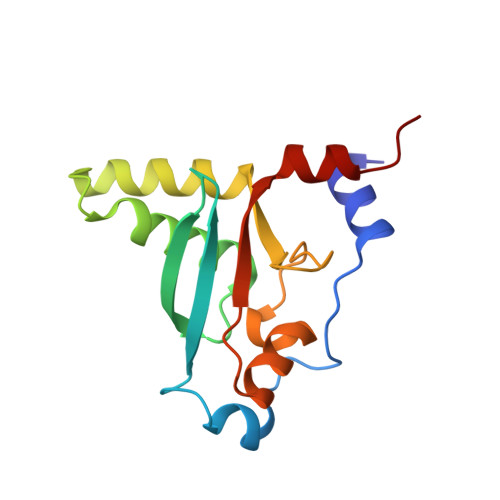
6VO7 - PubMed Abstract:
Phosphatidylinositol-3-kinases (PI3Ks) are lipid kinases that phosphorylate phosphatidylinositol 4,5-bisphosphate to generate a key lipid second messenger, phosphatidylinositol 3,4,5-bisphosphate. PI3Kα and PI3Kγ require activation by RAS proteins to stimulate signaling pathways that control cellular growth, differentiation, motility and survival. Intriguingly, RAS binding to PI3K isoforms likely differ, as RAS mutations have been identified that discriminate between PI3Kα and PI3Kγ, consistent with low sequence homology (23%) between their RAS binding domains (RBDs). As disruption of the RAS/PI3Kα interaction reduces tumor growth in mice with RAS- and epidermal growth factor receptor driven skin and lung cancers, compounds that interfere with this key interaction may prove useful as anti-cancer agents. However, a structure of PI3Kα bound to RAS is lacking, limiting drug discovery efforts. Expression of full-length PI3K isoforms in insect cells has resulted in low yield and variable activity, limiting biophysical and structural studies of RAS/PI3K interactions. This led us to generate the first RBDs from PI3Kα and PI3Kγ that can be expressed at high yield in bacteria and bind to RAS with similar affinity to full-length PI3K. We also solved a 2.31 Å X-ray crystal structure of the PI3Kα-RBD, which aligns well to full-length PI3Kα. Structural differences between the PI3Kα and PI3Kγ RBDs are consistent with differences in thermal stability and may underly differential RAS recognition and RAS-mediated PI3K activation. These high expression, functional PI3K RBDs will aid in interrogating RAS interactions and could aid in identifying inhibitors of this key interaction.
- Department of Biochemistry & Biophysics, University of North Carolina at Chapel Hill, United States.
Organizational Affiliation: