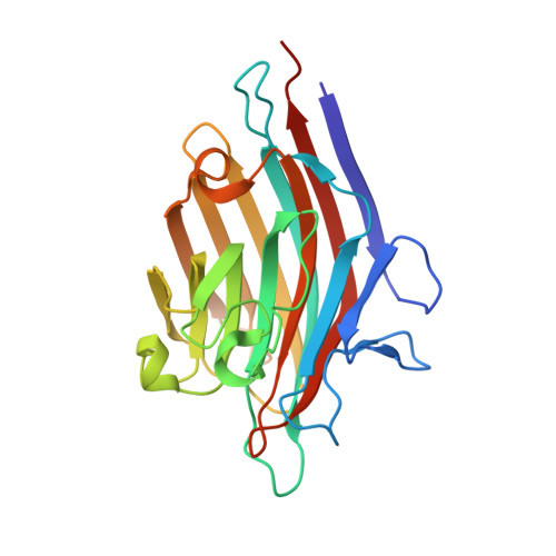
6VC3

Peanut lectin complexed with S-beta-D-thiogalactopyranosyl 6-deoxy-6-S-propynyl-beta-D-glucopyranoside (STG)


Experimental Data Snapshot

  • Method: X-RAY DIFFRACTION
  • Resolution: 1.95 Å
  • R-Value Free: 0.283 
  • R-Value Work: 0.267 
  • R-Value Observed: 0.268 

wwPDB Validation   3D Report Full Report

Currently 6VC3 does not have a validation slider image.


This is version 1.2 of the entry. See complete history


Literature

Crystal structures of peanut lectin in the presence of synthetic beta-N- and beta-S-galactosides disclose evidence for the recognition of different glycomimetic ligands.

Cagnoni, A.J.Primo, E.D.Klinke, S.Cano, M.E.Giordano, W.Marino, K.V.Kovensky, J.Goldbaum, F.A.Uhrig, M.L.Otero, L.H.

(2020) Acta Crystallogr D Struct Biol 76: 1080-1091

  • DOI: https://doi.org/10.1107/S2059798320012371
  • Primary Citation of Related Structures:  
    6V95, 6VAV, 6VAW, 6VC3, 6VC4, 6VGF

  • PubMed Abstract: 

    Carbohydrate-lectin interactions are involved in important cellular recognition processes, including viral and bacterial infections, inflammation and tumor metastasis. Hence, structural studies of lectin-synthetic glycan complexes are essential for understanding lectin-recognition processes and for the further design of promising chemotherapeutics that interfere with sugar-lectin interactions. Plant lectins are excellent models for the study of the molecular-recognition process. Among them, peanut lectin (PNA) is highly relevant in the field of glycobiology because of its specificity for β-galactosides, showing high affinity towards the Thomsen-Friedenreich antigen, a well known tumor-associated carbohydrate antigen. Given this specificity, PNA is one of the most frequently used molecular probes for the recognition of tumor cell-surface O-glycans. Thus, it has been extensively used in glycobiology for inhibition studies with a variety of β-galactoside and β-lactoside ligands. Here, crystal structures of PNA are reported in complex with six novel synthetic hydrolytically stable β-N- and β-S-galactosides. These complexes disclosed key molecular-binding interactions of the different sugars with PNA at the atomic level, revealing the roles of specific water molecules in protein-ligand recognition. Furthermore, binding-affinity studies by isothermal titration calorimetry showed dissociation-constant values in the micromolar range, as well as a positive multivalency effect in terms of affinity in the case of the divalent compounds. Taken together, this work provides a qualitative structural rationale for the upcoming synthesis of optimized glycoclusters designed for the study of lectin-mediated biological processes. The understanding of the recognition of β-N- and β-S-galactosides by PNA represents a benchmark in protein-carbohydrate interactions since they are novel synthetic ligands that do not belong to the family of O-linked glycosides.


  • Organizational Affiliation

    Laboratorio de Glicómica Funcional y Molecular, Instituto de Biología y Medicina Experimental, IBYME-CONICET, Vuelta de Obligado 2490, C1428ADN Buenos Aires, Argentina.


Macromolecules
Find similar proteins by:  (by identity cutoff)  |  3D Structure
Entity ID: 1
MoleculeChains Sequence LengthOrganismDetailsImage
Galactose-binding lectin
A, B, C, D
236Arachis hypogaeaMutation(s): 0 
UniProt
Find proteins for P02872 (Arachis hypogaea)
Explore P02872 
Go to UniProtKB:  P02872
Entity Groups  
Sequence Clusters30% Identity50% Identity70% Identity90% Identity95% Identity100% Identity
UniProt GroupP02872
Sequence Annotations
Expand
  • Reference Sequence
Small Molecules
Ligands 3 Unique
IDChains Name / Formula / InChI Key2D Diagram3D Interactions
QWJ (Subject of Investigation/LOI)
Query on QWJ

Download Ideal Coordinates CCD File 
E [auth A],
H [auth B],
M [auth D]
6-S-(prop-2-yn-1-yl)-6-thio-beta-D-glucopyranosyl 1-thio-beta-D-galactopyranoside
C15 H24 O9 S2
VMLLJSJIKUKSIE-PHOFNFIUSA-N
MN
Query on MN

Download Ideal Coordinates CCD File 
F [auth A],
I [auth B],
K [auth C],
N [auth D]
MANGANESE (II) ION
Mn
WAEMQWOKJMHJLA-UHFFFAOYSA-N
CA
Query on CA

Download Ideal Coordinates CCD File 
G [auth A],
J [auth B],
L [auth C],
O [auth D]
CALCIUM ION
Ca
BHPQYMZQTOCNFJ-UHFFFAOYSA-N
Binding Affinity Annotations 
IDSourceBinding Affinity
QWJ Binding MOAD:  6VC3 Kd: 4.82e+5 (nM) from 1 assay(s)
Experimental Data & Validation

Experimental Data

  • Method: X-RAY DIFFRACTION
  • Resolution: 1.95 Å
  • R-Value Free: 0.283 
  • R-Value Work: 0.267 
  • R-Value Observed: 0.268 
  • Space Group: P 2 21 21
Unit Cell:
Length ( Å )Angle ( ˚ )
a = 76.19α = 90
b = 125.418β = 90
c = 128.112γ = 90
Software Package:
Software NamePurpose
BUSTERrefinement
PDB_EXTRACTdata extraction
XDSdata reduction
Aimlessdata scaling
PHASERphasing
MxCuBEdata collection

Structure Validation

View Full Validation Report

Currently 6VC3 does not have a validation slider image.


Entry History & Funding Information

Deposition Data


Funding OrganizationLocationGrant Number
National Research Council (NRC, Argentina)ArgentinaPICT-2015-0621
National Research Council (NRC, Argentina)ArgentinaPICT-2016-1425

Revision History  (Full details and data files)

  • Version 1.0: 2020-10-28
    Type: Initial release
  • Version 1.1: 2020-11-18
    Changes: Database references
  • Version 1.2: 2023-10-11
    Changes: Advisory, Data collection, Database references, Refinement description