6V6U | pdb_00006v6u

Crystal structure of RhoA-GDP with novel Switch I conformation


Experimental Data Snapshot

  • Method: X-RAY DIFFRACTION
  • Resolution: 1.16 Å
  • R-Value Free: 
    0.185 (Depositor), 0.184 (DCC) 
  • R-Value Work: 
    0.170 (Depositor), 0.170 (DCC) 
  • R-Value Observed: 
    0.171 (Depositor) 

Starting Model: experimental
View more details

wwPDB Validation   3D Report Full Report


Ligand Structure Quality Assessment 


This is version 1.3 of the entry. See complete history


Literature

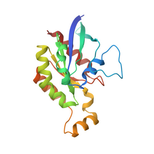
Structure of an inactive conformation of GTP-bound RhoA GTPase.

Lin, Y.Lu, S.Zhang, J.Zheng, Y.

(2021) Structure 29: 553-563.e5

  • DOI: https://doi.org/10.1016/j.str.2020.12.015
  • Primary Citation Related Structures: 
    6V6M, 6V6U, 6V6V

  • PubMed Abstract: 

    By using 31 P NMR, we present evidence that the Rho family GTPase RhoA, similar to Ras GTPases, exists in an equilibrium of conformations when bound to GTP. High-resolution crystal structures of RhoA bound to the GTP analog GMPPNP and to GDP show that they display a similar overall inactive conformation. In contrast to the previously reported crystal structures of GTP analog-bound forms of two RhoA dominantly active mutants (G14V and Q63L), GMPPNP-bound RhoA assumes an open conformation in the Switch I loop with a previously unseen interaction between the γ-phosphate and Pro36, instead of the canonical Thr37. Molecular dynamics simulations found that the oncogenic RhoA G14V mutant displays a reduced flexibility in the Switch regions, consistent with a crystal structure of GDP-bound RhoA G14V . Thus, GDP- and GTP-bound RhoA can present similar inactive conformations, and the molecular dynamics in the Switch regions are likely to have a role in RhoA activation.


  • Organizational Affiliation
    • Experimental Hematology and Cancer Biology, Cincinnati Children's Hospital Medical Center, University of Cincinnati College of Medicine, 3333 Burnet Avenue, Cincinnati, OH 45229, USA.

Macromolecules
Find similar proteins by:  (by identity cutoff)  |  3D Structure
Entity ID: 1
MoleculeChains Sequence LengthOrganismDetailsImage
Transforming protein RhoA183Homo sapiensMutation(s): 1 
Gene Names: RHOAARH12ARHARHO12
EC: 3.6.5.2
UniProt & NIH Common Fund Data Resources
Find proteins for P61586 (Homo sapiens)
Explore P61586 
Go to UniProtKB:  P61586
PHAROS:  P61586
GTEx:  ENSG00000067560 
Entity Groups  
Sequence Clusters30% Identity50% Identity70% Identity90% Identity95% Identity100% Identity
UniProt GroupP61586
Sequence Annotations
Expand
  • Reference Sequence
Experimental Data & Validation

Experimental Data

  • Method: X-RAY DIFFRACTION
  • Resolution: 1.16 Å
  • R-Value Free:  0.185 (Depositor), 0.184 (DCC) 
  • R-Value Work:  0.170 (Depositor), 0.170 (DCC) 
  • R-Value Observed: 0.171 (Depositor) 
Space Group: P 1 21 1
Unit Cell:
Length ( Å )Angle ( ˚ )
a = 32.216α = 90
b = 87.192β = 95.06
c = 34.271γ = 90
Software Package:
Software NamePurpose
PHENIXrefinement
MOSFLMdata reduction
SCALAdata scaling
PHASERphasing
PDB_EXTRACTdata extraction

Structure Validation

View Full Validation Report


Ligand Structure Quality Assessment 


Entry History 

Deposition Data

Revision History  (Full details and data files)

  • Version 1.0: 2020-12-23
    Type: Initial release
  • Version 1.1: 2021-02-10
    Changes: Database references
  • Version 1.2: 2021-06-16
    Changes: Database references
  • Version 1.3: 2023-10-11
    Changes: Data collection, Database references, Refinement description