Kinetic and structural analysis of Escherichia coli phosphoenolpyruvate carboxykinase mutants.
Sokaribo, A., Novakovski, B.A.A., Cotelesage, J., White, A.P., Sanders, D., Goldie, H.(2020) Biochim Biophys Acta Gen Subj 1864: 129517-129517
- PubMed: 31911238 Search on PubMed
- DOI: https://doi.org/10.1016/j.bbagen.2020.129517
- Primary Citation Related Structures:
6COM, 6V2L, 6V2M, 6V2N - PubMed Abstract:
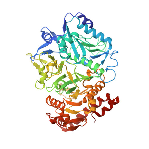
Phosphoenolpyruvate carboxykinase (PEPCK) is a metabolic enzyme in the gluconeogenesis pathway, where it catalyzes the reversible conversion of oxaloacetate (OAA) to phosphoenolpyruvate (PEP) and CO 2 . The substrates for Escherichia coli PEPCK are OAA and MgATP, with Mn 2+ acting as a cofactor. Analysis of PEPCK structures have revealed amino acid residues involved in substrate/cofactor coordination during catalysis. Key residues involved in coordinating the different substrates and cofactor bound to E. coli PEPCK were mutated. Purified mutant enzymes were used for kinetic assays. The structure of some mutant enzymes were determined using X-ray crystallography. Mutation of residues D269 and H232, which comprise part of the coordination sphere of Mn 2+ , reduced k cat by 14-fold, and significantly increased the K m values for Mn 2+ and OAA. Mutation of K254 a key residue in the P-loop motif that interacts with MgATP, significantly elevated the K m value for MgATP and reduced k cat . R65 and R333 are key residues that interacts with OAA. The R65Q and R333Q mutations significantly increased the K m value for OAA and reduced k cat respectively. Our results show that mutation of residues involved in coordinating OAA, MgATP and Mn 2+ significantly reduce PEPCK activity. K254 plays an important role in phosphoryl transfer, while R333 is involved in both OAA decarboxylation and phosphoryl transfer by E. coli PEPCK. In higher organisms including humans, PEPCK helps to regulate blood glucose levels, hence PEPCK is a potential drug target for patients with non-insulin dependent diabetes mellitus.
- Department of Biochemistry, Microbiology and Immunology, University of Saskatchewan, Saskatoon, SK, Canada; Vaccine and Infectious Disease Organization, International Vaccine Center, Saskatoon, SK, Canada. Electronic address: ats172@mail.usask.ca.
Organizational Affiliation: