6UYC | pdb_00006uyc

Crystal structure of TEAD2 bound to Compound 2


Experimental Data Snapshot

  • Method: X-RAY DIFFRACTION
  • Resolution: 1.66 Å
  • R-Value Free: 
    0.216 (Depositor), 0.219 (DCC) 
  • R-Value Work: 
    0.194 (Depositor), 0.196 (DCC) 
  • R-Value Observed: 
    0.195 (Depositor) 

Starting Model: experimental
View more details

wwPDB Validation 3D Report Full Report

Validation slider image for 6UYC

Ligand Structure Quality Assessment 


This is version 1.2 of the entry. See complete history

Literature

Small Molecule Dysregulation of TEAD Lipidation Induces a Dominant-Negative Inhibition of Hippo Pathway Signaling.

Holden, J.K.Crawford, J.J.Noland, C.L.Schmidt, S.Zbieg, J.R.Lacap, J.A.Zang, R.Miller, G.M.Zhang, Y.Beroza, P.Reja, R.Lee, W.Tom, J.Y.K.Fong, R.Steffek, M.Clausen, S.Hagenbeek, T.J.Hu, T.Zhou, Z.Shen, H.C.Cunningham, C.N.

(2020) Cell Rep 31: 107809-107809

  • DOI: https://doi.org/10.1016/j.celrep.2020.107809
  • Primary Citation Related Structures: 
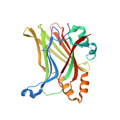
    6UYB, 6UYC

  • PubMed Abstract: 

    The transcriptional enhanced associate domain (TEAD) family of transcription factors serves as the receptors for the downstream effectors of the Hippo pathway, YAP and TAZ, to upregulate the expression of multiple genes involved in cellular proliferation and survival. Recent work identified TEAD S-palmitoylation as critical for protein stability and activity as the lipid tail extends into a hydrophobic core of the protein. Here, we report the identification and characterization of a potent small molecule that binds the TEAD lipid pocket (LP) and disrupts TEAD S-palmitoylation. Using a variety of biochemical, structural, and cellular methods, we uncover that TEAD S-palmitoylation functions as a TEAD homeostatic protein level checkpoint and that dysregulation of this lipidation affects TEAD transcriptional activity in a dominant-negative manner. Furthermore, we demonstrate that targeting the TEAD LP is a promising therapeutic strategy for modulating the Hippo pathway, showing tumor stasis in a mouse xenograft model.


  • Organizational Affiliation
    • Department of Early Discovery Biochemistry, Genentech, South San Francisco, CA 94080, USA.

Macromolecule Content 

  • Total Structure Weight: 54.22 kDa 
  • Atom Count: 3,523 
  • Modeled Residue Count: 404 
  • Deposited Residue Count: 472 
  • Unique protein chains: 1

Macromolecules

Find similar proteins by:|  3D Structure
Entity ID: 1
MoleculeChains  Sequence LengthOrganismDetailsImage
Transcriptional enhancer factor TEF-4
A, B
236Homo sapiensMutation(s): 0 
Gene Names: TEAD2TEF4
UniProt & NIH Common Fund Data Resources
Find proteins for Q15562 (Homo sapiens)
Explore Q15562 
Go to UniProtKB:  Q15562
PHAROS:  Q15562
GTEx:  ENSG00000074219 
Entity Groups
Sequence Clusters30% Identity50% Identity70% Identity90% Identity95% Identity100% Identity
UniProt GroupQ15562
Sequence Annotations
Expand
Reference Sequence

Small Molecules

Binding Affinity Annotations 
IDSourceBinding Affinity
QLV BindingDB:  6UYC Kd: 229 (nM) from 1 assay(s)

Experimental Data & Validation

Experimental Data

  • Method: X-RAY DIFFRACTION
  • Resolution: 1.66 Å
  • R-Value Free:  0.216 (Depositor), 0.219 (DCC) 
  • R-Value Work:  0.194 (Depositor), 0.196 (DCC) 
  • R-Value Observed: 0.195 (Depositor) 
Space Group: C 1 2 1
Unit Cell:
Length ( Å )Angle ( ˚ )
a = 123.422α = 90
b = 61.249β = 117.26
c = 80.206γ = 90
Software Package:
Software NamePurpose
PHENIXrefinement
PDB_EXTRACTdata extraction
ADDREFdata reduction
autoPROCdata scaling
PHENIXphasing

Structure Validation

View Full Validation Report


Ligand Structure Quality Assessment 


Entry History 

Deposition Data

Revision History  (Full details and data files)

  • Version 1.0: 2020-06-24
    Type: Initial release
  • Version 1.1: 2020-07-08
    Changes: Database references
  • Version 1.2: 2023-10-11
    Changes: Data collection, Database references, Refinement description