6UY8

Crystal structure of the STAC3 tandem SH3 domains - P269R, K329N


Experimental Data Snapshot

  • Method: X-RAY DIFFRACTION
  • Resolution: 1.65 Å
  • R-Value Free: 0.208 
  • R-Value Work: 0.187 
  • R-Value Observed: 0.189 

wwPDB Validation   3D Report Full Report

Currently 6UY8 does not have a validation slider image.


This is version 1.2 of the entry. See complete history


Literature

Multiple Sequence Variants in STAC3 Affect Interactions with CaV1.1 and Excitation-Contraction Coupling.

Rufenach, B.Christy, D.Flucher, B.E.Bui, J.M.Gsponer, J.Campiglio, M.Van Petegem, F.

(2020) Structure 28: 922

  • DOI: https://doi.org/10.1016/j.str.2020.05.005
  • Primary Citation of Related Structures:  
    6UY7, 6UY8, 6UY9

  • PubMed Abstract: 

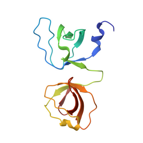
    STAC3 is a soluble protein essential for skeletal muscle excitation-contraction (EC) coupling. Through its tandem SH3 domains, it interacts with the cytosolic II-III loop of the skeletal muscle voltage-gated calcium channel. STAC3 is the target for a mutation (W284S) that causes Native American myopathy, but multiple other sequence variants have been reported. Here, we report a crystal structure of the human STAC3 tandem SH3 domains. We analyzed the effect of five disease-associated variants, spread over both SH3 domains, on their ability to bind to the Ca V 1.1 II-III loop and on muscle EC coupling. In addition to W284S, we find the F295L and K329N variants to affect both binding and EC coupling. The ability of the K329N variant, located in the second SH3 domain, to affect the interaction highlights the importance of both SH3 domains in association with Ca V 1.1. Our results suggest that multiple STAC3 variants may cause myopathy.


  • Organizational Affiliation

    Department of Biochemistry and Molecular Biology, University of British Columbia, Vancouver, BC V6T 1Z3, Canada.


Macromolecules
Find similar proteins by:  (by identity cutoff)  |  3D Structure
Entity ID: 1
MoleculeChains Sequence LengthOrganismDetailsImage
SH3 and cysteine-rich domain-containing protein 3
A, B
123Homo sapiensMutation(s): 2 
Gene Names: STAC3
UniProt & NIH Common Fund Data Resources
Find proteins for Q96MF2 (Homo sapiens)
Explore Q96MF2 
Go to UniProtKB:  Q96MF2
PHAROS:  Q96MF2
GTEx:  ENSG00000185482 
Entity Groups  
Sequence Clusters30% Identity50% Identity70% Identity90% Identity95% Identity100% Identity
UniProt GroupQ96MF2
Sequence Annotations
Expand
  • Reference Sequence
Experimental Data & Validation

Experimental Data

  • Method: X-RAY DIFFRACTION
  • Resolution: 1.65 Å
  • R-Value Free: 0.208 
  • R-Value Work: 0.187 
  • R-Value Observed: 0.189 
  • Space Group: P 32
Unit Cell:
Length ( Å )Angle ( ˚ )
a = 87.878α = 90
b = 87.878β = 90
c = 29.452γ = 120
Software Package:
Software NamePurpose
HKL-2000data scaling
PHENIXrefinement
PDB_EXTRACTdata extraction
HKL-2000data reduction
PHASERphasing

Structure Validation

View Full Validation Report

Currently 6UY8 does not have a validation slider image.


Entry History & Funding Information

Deposition Data


Funding OrganizationLocationGrant Number
Canadian Institutes of Health Research (CIHR)CanadaPJT-159601

Revision History  (Full details and data files)

  • Version 1.0: 2020-06-17
    Type: Initial release
  • Version 1.1: 2020-08-19
    Changes: Database references
  • Version 1.2: 2023-10-11
    Changes: Data collection, Database references, Refinement description