


Crystal structure of acetoin dehydrogenase from Enterobacter cloacae
Chang, C., Skarina, T., Mesa, N., Savchenko, A., Joachimiak, A., Center for Structural Genomics of Infectious Diseases (CSGID)To be published.
Experimental Data Snapshot
Starting Model: experimental
View more details
wwPDB Validation 3D Report Full Report

Entity ID: 1 | |||||
|---|---|---|---|---|---|
| Molecule | Chains | Sequence Length | Organism | Details | Image |
| Acetoin dehydrogenase | 257 | Enterobacter cloacae subsp. cloacae ATCC 13047 | Mutation(s): 0 Gene Names: ECL_03449 | 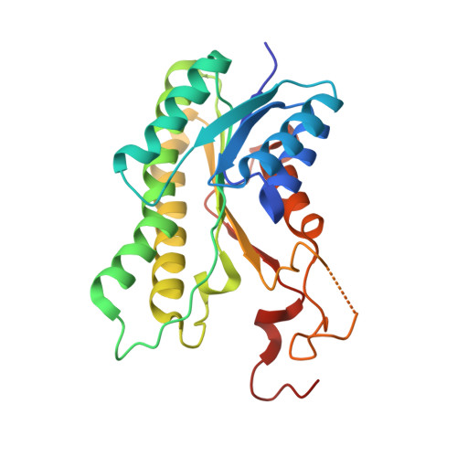 | |
UniProt | |||||
Find proteins for A0A0H3CM71 (Enterobacter cloacae subsp. cloacae (strain ATCC 13047 / DSM 30054 / NBRC 13535 / NCTC 10005 / WDCM 00083 / NCDC 279-56)) Explore A0A0H3CM71 Go to UniProtKB: A0A0H3CM71 | |||||
Entity Groups | |||||
| Sequence Clusters | 30% Identity50% Identity70% Identity90% Identity95% Identity100% Identity | ||||
| UniProt Group | A0A0H3CM71 | ||||
Sequence AnnotationsExpand | |||||
Reference Sequence | |||||
| Ligands 2 Unique | |||||
|---|---|---|---|---|---|
| ID | Chains | Name / Formula / InChI Key | 2D Diagram | 3D Interactions | |
| PEG Download:Ideal Coordinates CCD File | E [auth C] | DI(HYDROXYETHYL)ETHER C4 H10 O3 MTHSVFCYNBDYFN-UHFFFAOYSA-N |  | ||
| GOL Download:Ideal Coordinates CCD File | F [auth D] | GLYCEROL C3 H8 O3 PEDCQBHIVMGVHV-UHFFFAOYSA-N |  | ||
| Length ( Å ) | Angle ( ˚ ) |
|---|---|
| a = 64.07 | α = 90 |
| b = 99.836 | β = 98.89 |
| c = 82.217 | γ = 90 |
| Software Name | Purpose |
|---|---|
| PHENIX | refinement |
| SCALEPACK | data scaling |
| PDB_EXTRACT | data extraction |
| HKL-3000 | data reduction |
| HKL-3000 | phasing |


