6URM | pdb_00006urm

Crystal structure of vaccine-elicited receptor-binding site targeting antibody LPAF-a.01 in complex with Hemagglutinin H1 A/California/04/2009


Experimental Data Snapshot

  • Method: X-RAY DIFFRACTION
  • Resolution: 2.65 Å
  • R-Value Free: 
    0.262 (Depositor), 0.263 (DCC) 
  • R-Value Work: 
    0.215 (Depositor), 0.215 (DCC) 
  • R-Value Observed: 
    0.217 (Depositor) 

Starting Model: experimental
View more details

wwPDB Validation 3D Report Full Report

Validation slider image for 6URM

Ligand Structure Quality Assessment 


This is version 1.2 of the entry. See complete history

Literature

Identification and Structure of a Multidonor Class of Head-Directed Influenza-Neutralizing Antibodies Reveal the Mechanism for Its Recurrent Elicitation.

Cheung, C.S.Fruehwirth, A.Paparoditis, P.C.G.Shen, C.H.Foglierini, M.Joyce, M.G.Leung, K.Piccoli, L.Rawi, R.Silacci-Fregni, C.Tsybovsky, Y.Verardi, R.Wang, L.Wang, S.Yang, E.S.Zhang, B.Zhang, Y.Chuang, G.Y.Corti, D.Mascola, J.R.Shapiro, L.Kwong, P.D.Lanzavecchia, A.Zhou, T.

(2020) Cell Rep 32: 108088-108088

  • DOI: https://doi.org/10.1016/j.celrep.2020.108088
  • Primary Citation Related Structures: 
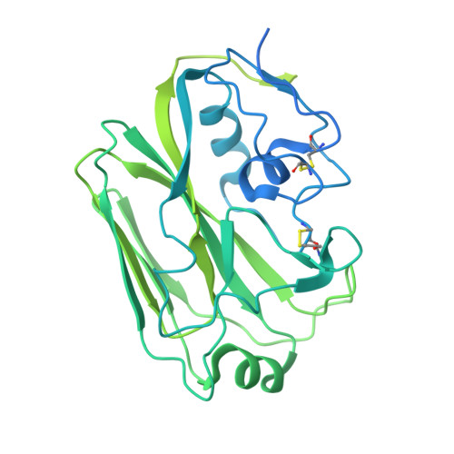
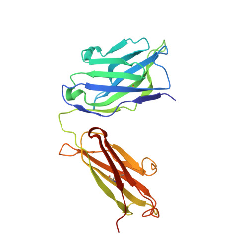
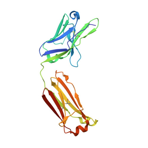
    6URM

  • PubMed Abstract: 

    Multidonor antibodies are of interest for vaccine design because they can in principle be elicited in the general population by a common set of immunogens. For influenza, multidonor antibodies have been observed against the hemagglutinin (HA) stem, but not the immunodominant HA head. Here, we identify and characterize a multidonor antibody class (LPAF-a class) targeting the HA head. This class exhibits potent viral entry inhibition against H1N1 A/California/04/2009 (CA09) virus. LPAF-a class antibodies derive from the HV2-70 gene and contain a "Tyr-Gly-Asp"-motif, which occludes the HA-sialic acid binding site as revealed by a co-crystal structure with HA. Both germline-reverted and mature LPAF antibodies potently neutralize CA09 virus and have nanomolar affinities for CA09 HA. Moreover, increased frequencies for LPFA-a class antibodies are observed in humans after a single vaccination. Overall, this work highlights the identification of a multidonor class of head-directed influenza-neutralizing antibodies and delineates the mechanism of their recurrent elicitation in humans.


  • Organizational Affiliation
    • Vaccine Research Center, National Institute of Allergy and Infectious Diseases, National Institutes of Health, Bethesda, MD 20892, USA.

Macromolecule Content 

  • Total Structure Weight: 209.01 kDa 
  • Atom Count: 10,193 
  • Modeled Residue Count: 1,303 
  • Deposited Residue Count: 1,886 
  • Unique protein chains: 3

Macromolecules

Find similar proteins by:|  3D Structure
Entity ID: 1
MoleculeChains  Sequence LengthOrganismDetailsImage
HemagglutininA [auth F],
D [auth C]
505Influenza A virusMutation(s): 0 
Gene Names: HA
UniProt
Find proteins for C3W5S1 (Influenza A virus (strain swl A/California/04/2009 H1N1))
Explore C3W5S1 
Go to UniProtKB:  C3W5S1
Entity Groups
Sequence Clusters30% Identity50% Identity70% Identity90% Identity95% Identity100% Identity
UniProt GroupC3W5S1
Glycosylation
Glycosylation Sites: 1
Sequence Annotations
Expand
Reference Sequence
Find similar proteins by:|  3D Structure
Entity ID: 2
MoleculeChains  Sequence LengthOrganismDetailsImage
The heavy chain of antibody LPAF-a.01B [auth H],
E [auth D]
227Homo sapiensMutation(s): 0 
Find similar proteins by:|  3D Structure
Entity ID: 3
MoleculeChains  Sequence LengthOrganismDetailsImage
The light chain of antibody LPAF-a.01C [auth L],
F [auth E]
211Homo sapiensMutation(s): 0 

Oligosaccharides

Help  
Entity ID: 4
MoleculeChains Length2D Diagram GlycosylationD Interactions
2-acetamido-2-deoxy-beta-D-glucopyranose-(1-4)-2-acetamido-2-deoxy-beta-D-glucopyranoseG [auth A]2N-Glycosylation
Glycosylation Resources
GlyTouCan: G42666HT
GlyCosmos: G42666HT
GlyGen: G42666HT
Entity ID: 5
MoleculeChains Length2D Diagram GlycosylationD Interactions
alpha-D-mannopyranose-(1-4)-2-acetamido-2-deoxy-beta-D-glucopyranose-(1-4)-2-acetamido-2-deoxy-beta-D-glucopyranoseH [auth B]3N-Glycosylation
Glycosylation Resources
GlyTouCan: G62182OO
GlyCosmos: G62182OO
GlyGen: G62182OO

Small Molecules

Ligands 3 Unique
IDChains Name / Formula / InChI Key2D Diagram3D Interactions
NAG

Query on NAG


Download:Ideal Coordinates CCD File
I [auth F]2-acetamido-2-deoxy-beta-D-glucopyranose
C8 H15 N O6
OVRNDRQMDRJTHS-FMDGEEDCSA-N
SO4

Query on SO4


Download:Ideal Coordinates CCD File
J [auth F],
L,
N [auth D],
O [auth E]
SULFATE ION
O4 S
QAOWNCQODCNURD-UHFFFAOYSA-L
GOL

Query on GOL


Download:Ideal Coordinates CCD File
K [auth H],
M [auth D]
GLYCEROL
C3 H8 O3
PEDCQBHIVMGVHV-UHFFFAOYSA-N

Experimental Data & Validation

Experimental Data

  • Method: X-RAY DIFFRACTION
  • Resolution: 2.65 Å
  • R-Value Free:  0.262 (Depositor), 0.263 (DCC) 
  • R-Value Work:  0.215 (Depositor), 0.215 (DCC) 
  • R-Value Observed: 0.217 (Depositor) 
Space Group: P 1 21 1
Unit Cell:
Length ( Å )Angle ( ˚ )
a = 42.408α = 90
b = 260.836β = 96.93
c = 66.453γ = 90
Software Package:
Software NamePurpose
PHENIXrefinement
HKL-2000data reduction
SCALEPACKdata scaling
PHASERphasing
PDB_EXTRACTdata extraction

Structure Validation

View Full Validation Report


Ligand Structure Quality Assessment 


Entry History 

Deposition Data

Revision History  (Full details and data files)

  • Version 1.0: 2020-09-16
    Type: Initial release
  • Version 1.1: 2023-10-11
    Changes: Data collection, Database references, Refinement description
  • Version 1.2: 2024-11-06
    Changes: Structure summary