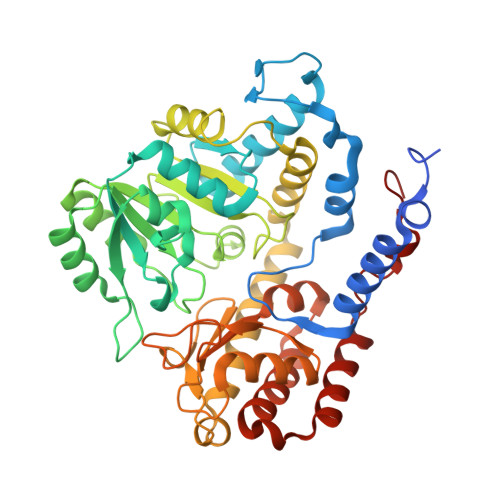
Crystal structure of serine hydroxymethyltransferase from Mycobacterium tuberculosis with bound PLP forming a Schiff base with substrate Serine in one monomer and PLP forming a Schiff base with product Glycine in the other monomer
Dranow, D.M., Abendroth, J., Lorimer, D.D., Horanyi, P.S., Edwards, T.E.To be published.