6UKO

Structure analysis of full-length mouse bcs1 complex


Experimental Data Snapshot

  • Method: X-RAY DIFFRACTION
  • Resolution: 4.40 Å
  • R-Value Free: 0.407 
  • R-Value Work: 0.357 
  • R-Value Observed: 0.358 

wwPDB Validation   3D Report Full Report


This is version 1.3 of the entry. See complete history


Literature

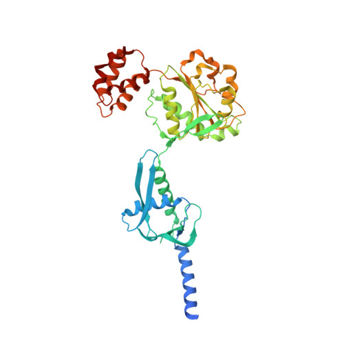
Structures of AAA protein translocase Bcs1 suggest translocation mechanism of a folded protein.

Tang, W.K.Borgnia, M.J.Hsu, A.L.Esser, L.Fox, T.de Val, N.Xia, D.

(2020) Nat Struct Mol Biol 27: 202-209

  • DOI: https://doi.org/10.1038/s41594-020-0373-0
  • Primary Citation of Related Structures:  
    6U1Y, 6UKO, 6UKP, 6UKS

  • PubMed Abstract: 

    The mitochondrial membrane-bound AAA protein Bcs1 translocate substrates across the mitochondrial inner membrane without previous unfolding. One substrate of Bcs1 is the iron-sulfur protein (ISP), a subunit of the respiratory Complex III. How Bcs1 translocates ISP across the membrane is unknown. Here we report structures of mouse Bcs1 in two different conformations, representing three nucleotide states. The apo and ADP-bound structures reveal a homo-heptamer and show a large putative substrate-binding cavity accessible to the matrix space. ATP binding drives a contraction of the cavity by concerted motion of the ATPase domains, which could push substrate across the membrane. Our findings shed light on the potential mechanism of translocating folded proteins across a membrane, offer insights into the assembly process of Complex III and allow mapping of human disease-associated mutations onto the Bcs1 structure.


  • Organizational Affiliation

    Laboratory of Cell Biology, National Cancer Institute, National Institutes of Health, Bethesda, MD, USA.


Macromolecules
Find similar proteins by:  (by identity cutoff)  |  3D Structure
Entity ID: 1
MoleculeChains Sequence LengthOrganismDetailsImage
Mitochondrial chaperone BCS1
A, B, C, D, E
A, B, C, D, E, F, G
424Mus musculusMutation(s): 0 
Gene Names: Bcs1l
Membrane Entity: Yes 
UniProt & NIH Common Fund Data Resources
Find proteins for Q9CZP5 (Mus musculus)
Explore Q9CZP5 
Go to UniProtKB:  Q9CZP5
IMPC:  MGI:1914071
Entity Groups  
Sequence Clusters30% Identity50% Identity70% Identity90% Identity95% Identity100% Identity
UniProt GroupQ9CZP5
Sequence Annotations
Expand
  • Reference Sequence
Small Molecules
Ligands 2 Unique
IDChains Name / Formula / InChI Key2D Diagram3D Interactions
ADP (Subject of Investigation/LOI)
Query on ADP

Download Ideal Coordinates CCD File 
H [auth A]
J [auth B]
L [auth C]
N [auth D]
P [auth E]
H [auth A],
J [auth B],
L [auth C],
N [auth D],
P [auth E],
R [auth F],
T [auth G]
ADENOSINE-5'-DIPHOSPHATE
C10 H15 N5 O10 P2
XTWYTFMLZFPYCI-KQYNXXCUSA-N
MG
Query on MG

Download Ideal Coordinates CCD File 
I [auth A]
K [auth B]
M [auth C]
O [auth D]
Q [auth E]
I [auth A],
K [auth B],
M [auth C],
O [auth D],
Q [auth E],
S [auth F],
U [auth G]
MAGNESIUM ION
Mg
JLVVSXFLKOJNIY-UHFFFAOYSA-N
Experimental Data & Validation

Experimental Data

  • Method: X-RAY DIFFRACTION
  • Resolution: 4.40 Å
  • R-Value Free: 0.407 
  • R-Value Work: 0.357 
  • R-Value Observed: 0.358 
  • Space Group: C 1 2 1
Unit Cell:
Length ( Å )Angle ( ˚ )
a = 254.055α = 90
b = 161.127β = 107.268
c = 132.595γ = 90
Software Package:
Software NamePurpose
PHENIXrefinement
HKL-2000data reduction
HKL-2000data scaling
MOLREPphasing

Structure Validation

View Full Validation Report


Entry History 

Deposition Data

Revision History  (Full details and data files)

  • Version 1.0: 2020-02-12
    Type: Initial release
  • Version 1.1: 2020-02-26
    Changes: Database references
  • Version 1.2: 2024-03-13
    Changes: Data collection, Database references, Refinement description
  • Version 1.3: 2024-04-03
    Changes: Refinement description