6UKL | pdb_00006ukl

Crystal Structure of a DiB2-split Protein


Experimental Data Snapshot

  • Method: X-RAY DIFFRACTION
  • Resolution: 2.02 Å
  • R-Value Free: 
    0.248 (Depositor), 0.254 (DCC) 
  • R-Value Work: 
    0.205 (Depositor), 0.214 (DCC) 
  • R-Value Observed: 
    0.207 (Depositor) 

Starting Model: experimental
View more details

wwPDB Validation 3D Report Full Report

Validation slider image for 6UKL

Ligand Structure Quality Assessment 


This is version 1.2 of the entry. See complete history

Literature

DiB-splits: nature-guided design of a novel fluorescent labeling split system.

Bozhanova, N.G.Gavrikov, A.S.Mishin, A.S.Meiler, J.

(2020) Sci Rep 10: 11049-11049

  • DOI: https://doi.org/10.1038/s41598-020-67095-2
  • Primary Citation Related Structures: 
    6UKK, 6UKL

  • PubMed Abstract: 

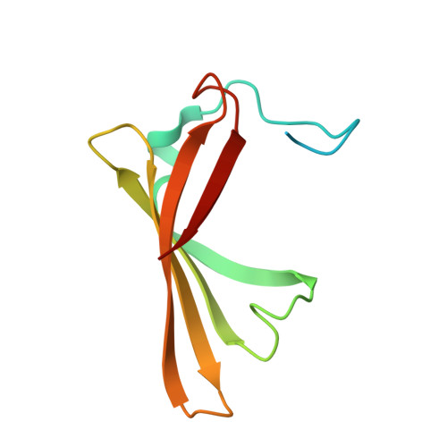
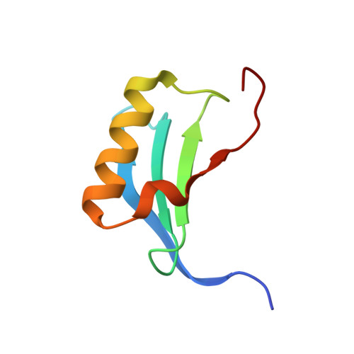
    Fluorogen-activating proteins (FAPs) are innovative fluorescent probes combining advantages of genetically-encoded proteins such as green fluorescent protein and externally added fluorogens that allow for highly tunable and on demand fluorescent signaling. Previously, a panel of green- and red-emitting FAPs has been created from bacterial lipocalin Blc (named DiBs). Here we present a rational design as well as functional and structural characterization of the first self-assembling FAP split system, DiB-splits. This new system decreases the size of the FAP label to ~8-12 kDa while preserving DiBs' unique properties: strong increase in fluorescence intensity of the chromophore upon binding, binding affinities to the chromophore in nanomolar to low micromolar range, and high photostability of the protein-ligand complex. These properties allow for use of DiB-splits for wide-field, confocal, and super-resolution fluorescence microscopy. DiB-splits also represent an attractive starting point for further design of a protein-protein interaction detection system as well as novel FAP-based sensors.


  • Organizational Affiliation
    • Department of Chemistry, Center for Structural Biology, Vanderbilt University, Nashville, TN, 37235, USA.

Macromolecule Content 

  • Total Structure Weight: 63.05 kDa 
  • Atom Count: 3,949 
  • Modeled Residue Count: 457 
  • Deposited Residue Count: 528 
  • Unique protein chains: 2

Macromolecules

Find similar proteins by:|  3D Structure
Entity ID: 1
MoleculeChains  Sequence LengthOrganismDetailsImage
Outer membrane lipoprotein BlcA,
B [auth C],
C [auth E]
107Escherichia coliMutation(s): 1 
Gene Names: blcZ5756ECs5130
UniProt
Find proteins for P0A901 (Escherichia coli (strain K12))
Explore P0A901 
Go to UniProtKB:  P0A901
Entity Groups
Sequence Clusters30% Identity50% Identity70% Identity90% Identity95% Identity100% Identity
UniProt GroupP0A901
Sequence Annotations
Expand
Reference Sequence
Find similar proteins by:|  3D Structure
Entity ID: 2
MoleculeChains  Sequence LengthOrganismDetailsImage
Outer membrane lipoprotein BlcD,
E [auth F],
F [auth B]
69Escherichia coliMutation(s): 0 
Gene Names: blcZ5756ECs5130
UniProt
Find proteins for P0A901 (Escherichia coli (strain K12))
Explore P0A901 
Go to UniProtKB:  P0A901
Entity Groups
Sequence Clusters30% Identity50% Identity70% Identity90% Identity95% Identity100% Identity
UniProt GroupP0A901
Sequence Annotations
Expand
Reference Sequence

Small Molecules

Ligands 3 Unique
IDChains Name / Formula / InChI Key2D Diagram3D Interactions
HEM

Query on HEM


Download:Ideal Coordinates CCD File
R [auth E]PROTOPORPHYRIN IX CONTAINING FE
C34 H32 Fe N4 O4
KABFMIBPWCXCRK-RGGAHWMASA-L
MES

Query on MES


Download:Ideal Coordinates CCD File
O [auth C],
U [auth D]
2-(N-MORPHOLINO)-ETHANESULFONIC ACID
C6 H13 N O4 S
SXGZJKUKBWWHRA-UHFFFAOYSA-N
SO4

Query on SO4


Download:Ideal Coordinates CCD File
G [auth A]
H [auth A]
I [auth A]
J [auth A]
K [auth C]
G [auth A],
H [auth A],
I [auth A],
J [auth A],
K [auth C],
L [auth C],
M [auth C],
N [auth C],
P [auth E],
Q [auth E],
S [auth D],
T [auth D],
V [auth B],
W [auth B],
X [auth B]
SULFATE ION
O4 S
QAOWNCQODCNURD-UHFFFAOYSA-L

Experimental Data & Validation

Experimental Data

  • Method: X-RAY DIFFRACTION
  • Resolution: 2.02 Å
  • R-Value Free:  0.248 (Depositor), 0.254 (DCC) 
  • R-Value Work:  0.205 (Depositor), 0.214 (DCC) 
  • R-Value Observed: 0.207 (Depositor) 
Space Group: P 32 2 1
Unit Cell:
Length ( Å )Angle ( ˚ )
a = 68.241α = 90
b = 68.241β = 90
c = 216.292γ = 120
Software Package:
Software NamePurpose
REFMACrefinement
xia2data scaling
MOLREPphasing
PDB_EXTRACTdata extraction
xia2data reduction

Structure Validation

View Full Validation Report


Ligand Structure Quality Assessment 


Entry History 

& Funding Information

Deposition Data


Funding OrganizationLocationGrant Number
National Institutes of Health/National Institute of General Medical Sciences (NIH/NIGMS)United StatesR01 GM099842

Revision History  (Full details and data files)

  • Version 1.0: 2020-07-08
    Type: Initial release
  • Version 1.1: 2020-07-22
    Changes: Database references
  • Version 1.2: 2023-10-11
    Changes: Data collection, Database references, Refinement description