6UE6 | pdb_00006ue6

PWWP1 domain of NSD2 in complex with MR837


Experimental Data Snapshot

  • Method: X-RAY DIFFRACTION
  • Resolution: 2.40 Å
  • R-Value Free: 
    0.252 (Depositor), 0.273 (DCC) 
  • R-Value Work: 
    0.235 (Depositor), 0.248 (DCC) 
  • R-Value Observed: 
    0.236 (Depositor) 

Starting Model: experimental
View more details

wwPDB Validation 3D Report Full Report

Validation slider image for 6UE6

Ligand Structure Quality Assessment 


This is version 1.2 of the entry. See complete history

Literature

Discovery of Small-Molecule Antagonists of the PWWP Domain of NSD2.

Ferreira de Freitas, R.Liu, Y.Szewczyk, M.M.Mehta, N.Li, F.McLeod, D.Zepeda-Velazquez, C.Dilworth, D.Hanley, R.P.Gibson, E.Brown, P.J.Al-Awar, R.James, L.I.Arrowsmith, C.H.Barsyte-Lovejoy, D.Min, J.Vedadi, M.Schapira, M.Allali-Hassani, A.

(2021) J Med Chem 64: 1584-1592

  • DOI: https://doi.org/10.1021/acs.jmedchem.0c01768
  • Primary Citation Related Structures: 
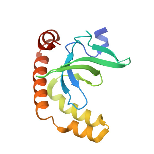
    6UE6

  • PubMed Abstract: 

    Increased activity of the lysine methyltransferase NSD2 driven by translocation and activating mutations is associated with multiple myeloma and acute lymphoblastic leukemia, but no NSD2-targeting chemical probe has been reported to date. Here, we present the first antagonists that block the protein-protein interaction between the N-terminal PWWP domain of NSD2 and H3K36me2. Using virtual screening and experimental validation, we identified the small-molecule antagonist 3f , which binds to the NSD2-PWWP1 domain with a K d of 3.4 μM and abrogates histone H3K36me2 binding to the PWWP1 domain in cells. This study establishes an alternative approach to targeting NSD2 and provides a small-molecule antagonist that can be further optimized into a chemical probe to better understand the cellular function of this protein.


  • Organizational Affiliation
    • Structural Genomics Consortium, University of Toronto, Toronto, Ontario M5G 1L7, Canada.

Macromolecule Content 

  • Total Structure Weight: 130.79 kDa 
  • Atom Count: 7,347 
  • Modeled Residue Count: 966 
  • Deposited Residue Count: 1,128 
  • Unique protein chains: 1

Macromolecules

Find similar proteins by:|  3D Structure
Entity ID: 1
MoleculeChains  Sequence LengthOrganismDetailsImage
Histone-lysine N-methyltransferase NSD2
A, B, C, D, E
A, B, C, D, E, F, G, H
141Homo sapiensMutation(s): 0 
Gene Names: NSD2KIAA1090MMSETTRX5WHSC1
EC: 2.1.1.43 (PDB Primary Data), 2.1.1.357 (UniProt)
UniProt & NIH Common Fund Data Resources
Find proteins for O96028 (Homo sapiens)
Explore O96028 
Go to UniProtKB:  O96028
PHAROS:  O96028
GTEx:  ENSG00000109685 
Entity Groups
Sequence Clusters30% Identity50% Identity70% Identity90% Identity95% Identity100% Identity
UniProt GroupO96028
Sequence Annotations
Expand
Reference Sequence

Small Molecules

Ligands 2 Unique
IDChains Name / Formula / InChI Key2D Diagram3D Interactions
Q5D
(Subject of Investigation/LOI)

Query on Q5D


Download:Ideal Coordinates CCD File
EA [auth C]
I [auth A]
KA [auth D]
LB [auth H]
OA [auth E]
EA [auth C],
I [auth A],
KA [auth D],
LB [auth H],
OA [auth E],
S [auth B],
UA [auth F],
ZA [auth G]
4-cyano-N-cyclopropyl-N-[(thiophen-2-yl)methyl]benzamide
C16 H14 N2 O S
WYAGGMZTARRIDX-UHFFFAOYSA-N
UNX

Query on UNX


Download:Ideal Coordinates CCD File
AA [auth B]
AB [auth G]
BA [auth B]
BB [auth G]
CA [auth B]
AA [auth B],
AB [auth G],
BA [auth B],
BB [auth G],
CA [auth B],
CB [auth G],
DA [auth B],
DB [auth G],
EB [auth G],
FA [auth C],
FB [auth G],
GA [auth C],
GB [auth G],
HA [auth C],
HB [auth G],
IA [auth C],
IB [auth G],
J [auth A],
JA [auth C],
JB [auth G],
K [auth A],
KB [auth G],
L [auth A],
LA [auth D],
M [auth A],
MA [auth D],
MB [auth H],
N [auth A],
NA [auth D],
NB [auth H],
O [auth A],
OB [auth H],
P [auth A],
PA [auth E],
Q [auth A],
QA [auth E],
R [auth A],
RA [auth E],
SA [auth E],
T [auth B],
TA [auth E],
U [auth B],
V [auth B],
VA [auth F],
W [auth B],
WA [auth F],
X [auth B],
XA [auth F],
Y [auth B],
YA [auth F],
Z [auth B]
UNKNOWN ATOM OR ION
X
Binding Affinity Annotations 
IDSourceBinding Affinity
Q5D BindingDB:  6UE6 Kd: min: 349, max: 7000 (nM) from 3 assay(s)
IC50: min: 1.73e+4, max: 2.47e+4 (nM) from 2 assay(s)

Experimental Data & Validation

Experimental Data

  • Method: X-RAY DIFFRACTION
  • Resolution: 2.40 Å
  • R-Value Free:  0.252 (Depositor), 0.273 (DCC) 
  • R-Value Work:  0.235 (Depositor), 0.248 (DCC) 
  • R-Value Observed: 0.236 (Depositor) 
Space Group: P 21 21 21
Unit Cell:
Length ( Å )Angle ( ˚ )
a = 69.21α = 90
b = 70.32β = 90
c = 228.83γ = 90
Software Package:
Software NamePurpose
BUSTERrefinement
XDSdata reduction
PHASERphasing
XDSdata scaling

Structure Validation

View Full Validation Report


Ligand Structure Quality Assessment 


Entry History 

Revision History  (Full details and data files)

  • Version 1.0: 2019-11-13
    Type: Initial release
  • Version 1.1: 2021-04-21
    Changes: Database references
  • Version 1.2: 2023-10-11
    Changes: Data collection, Database references, Refinement description