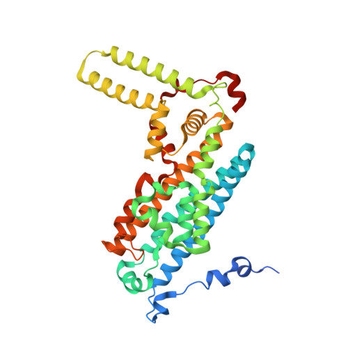
6UD5

Crystal structure of human tryptophan 2,3-dioxygenase in complex with carbon monoxide and tryptophan


Experimental Data Snapshot

  • Method: X-RAY DIFFRACTION
  • Resolution: 2.05 Å
  • R-Value Free: 0.217 
  • R-Value Work: 0.183 
  • R-Value Observed: 0.185 

wwPDB Validation   3D Report Full Report

Currently 6UD5 does not have a validation slider image.


This is version 1.2 of the entry. See complete history


Literature

Conformational Plasticity in Human Heme-Based Dioxygenases.

Pham, K.N.Lewis-Ballester, A.Yeh, S.R.

(2021) J Am Chem Soc 143: 1836-1845

  • DOI: https://doi.org/10.1021/jacs.0c09970
  • Primary Citation of Related Structures:  
    6UBP, 6UD5

  • PubMed Abstract: 

    Human indoleamine 2,3-dioxygenase 1 (hIDO1) and human tryptophan dioxygenase (hTDO) are two important heme proteins that degrade the essential amino acid, l-tryptophan (Trp), along the kynurenine pathway. The two enzymes share a similar active site structure and an analogous catalytic mechanism, but they exhibit a variety of distinct functional properties. Here we used carbon monoxide (CO) as a structural probe to interrogate how the functionalities of the two enzymes are encoded in their structures. With X-ray crystallography, we detected an unexpected photochemical intermediate trapped in a crystal of the hIDO1-CO-Trp complex, where CO is photolyzed from the heme iron by X-rays at cryogenic temperatures (100 K). The CO photolysis triggers a large-scale migration of the substrate Trp, as well as the photolyzed CO, from the active site to a temporary binding site, Sa*. It is accompanied by a large conformational change to an active site loop, JK-Loop C , despite the severely restricted protein motion under the frozen conditions, which highlights the remarkable conformational plasticity of the hIDO1 protein. Comparative studies of a crystal of the hTDO-CO-Trp complex show that CO and Trp remain bound in the active site under comparable X-ray illumination, indicating a much more rigid protein architecture. The data offer important new insights into the structure and function relationships of the heme-based dioxygenases and provide new guidelines for structure-based design of inhibitors targeting them.


  • Organizational Affiliation

    Department of Physiology and Biophysics, Albert Einstein College of Medicine, The Bronx, New York 10461, United States.


Macromolecules
Find similar proteins by:  (by identity cutoff)  |  3D Structure
Entity ID: 1
MoleculeChains Sequence LengthOrganismDetailsImage
Tryptophan 2,3-dioxygenase
A, B, C, D
380Homo sapiensMutation(s): 0 
Gene Names: TDO2TDO
EC: 1.13.11.11
UniProt & NIH Common Fund Data Resources
Find proteins for P48775 (Homo sapiens)
Explore P48775 
Go to UniProtKB:  P48775
PHAROS:  P48775
GTEx:  ENSG00000151790 
Entity Groups  
Sequence Clusters30% Identity50% Identity70% Identity90% Identity95% Identity100% Identity
UniProt GroupP48775
Sequence Annotations
Expand
  • Reference Sequence
Small Molecules
Ligands 3 Unique
IDChains Name / Formula / InChI Key2D Diagram3D Interactions
HEM
Query on HEM

Download Ideal Coordinates CCD File 
E [auth A],
I [auth B],
M [auth C],
Q [auth D]
PROTOPORPHYRIN IX CONTAINING FE
C34 H32 Fe N4 O4
KABFMIBPWCXCRK-RGGAHWMASA-L
TRP (Subject of Investigation/LOI)
Query on TRP

Download Ideal Coordinates CCD File 
G [auth A]
H [auth A]
K [auth B]
L [auth B]
O [auth C]
G [auth A],
H [auth A],
K [auth B],
L [auth B],
O [auth C],
P [auth C],
S [auth D],
T [auth D]
TRYPTOPHAN
C11 H12 N2 O2
QIVBCDIJIAJPQS-VIFPVBQESA-N
CMO
Query on CMO

Download Ideal Coordinates CCD File 
F [auth A],
J [auth B],
N [auth C],
R [auth D]
CARBON MONOXIDE
C O
UGFAIRIUMAVXCW-UHFFFAOYSA-N
Experimental Data & Validation

Experimental Data

  • Method: X-RAY DIFFRACTION
  • Resolution: 2.05 Å
  • R-Value Free: 0.217 
  • R-Value Work: 0.183 
  • R-Value Observed: 0.185 
  • Space Group: P 21 21 2
Unit Cell:
Length ( Å )Angle ( ˚ )
a = 144.725α = 90
b = 154.493β = 90
c = 88.306γ = 90
Software Package:
Software NamePurpose
REFMACrefinement
PDB_EXTRACTdata extraction
XDSdata reduction
SCALAdata scaling
PHASERphasing

Structure Validation

View Full Validation Report

Currently 6UD5 does not have a validation slider image.


Entry History & Funding Information

Deposition Data


Funding OrganizationLocationGrant Number
National Institutes of Health/National Institute of General Medical Sciences (NIH/NIGMS)United StatesGM115773
National Science Foundation (NSF, United States)United StatesCHE-1404929

Revision History  (Full details and data files)

  • Version 1.0: 2021-02-03
    Type: Initial release
  • Version 1.1: 2021-02-17
    Changes: Database references
  • Version 1.2: 2023-10-11
    Changes: Data collection, Database references, Refinement description