


Crystal structure of human PACRG in complex with MEIG1
Khan, N., Pelletier, D., Veyron, S., Croteau, N., Ichikawa, M., Black, C., Khalifa, A.A.Z., Chaaban, S., Kurinov, I., Brouhard, G., Bui, K.H., Trempe, J.F.(2019) bioRxiv
Experimental Data Snapshot
Starting Model: experimental
View more details
wwPDB Validation 3D Report Full Report

(2019) bioRxiv
Entity ID: 1 | |||||
|---|---|---|---|---|---|
| Molecule | Chains | Sequence Length | Organism | Details | Image |
| Parkin coregulated gene protein | 257 | Homo sapiens | Mutation(s): 0 Gene Names: PACRG, GLUP | 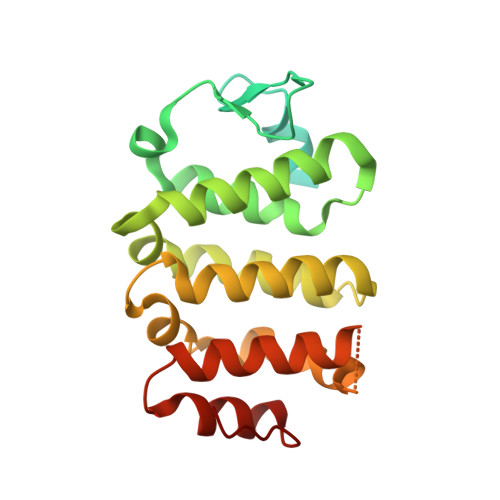 | |
UniProt & NIH Common Fund Data Resources | |||||
PHAROS: Q96M98 GTEx: ENSG00000112530 | |||||
Entity Groups | |||||
| Sequence Clusters | 30% Identity50% Identity70% Identity90% Identity95% Identity100% Identity | ||||
| UniProt Group | Q96M98 | ||||
Sequence AnnotationsExpand | |||||
Reference Sequence | |||||
Entity ID: 2 | |||||
|---|---|---|---|---|---|
| Molecule | Chains | Sequence Length | Organism | Details | Image |
| Meiosis expressed gene 1 protein homolog | 93 | Homo sapiens | Mutation(s): 0 Gene Names: MEIG1 | 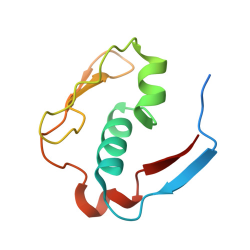 | |
UniProt & NIH Common Fund Data Resources | |||||
PHAROS: Q5JSS6 GTEx: ENSG00000197889 | |||||
Entity Groups | |||||
| Sequence Clusters | 30% Identity50% Identity70% Identity90% Identity95% Identity100% Identity | ||||
| UniProt Group | Q5JSS6 | ||||
Sequence AnnotationsExpand | |||||
Reference Sequence | |||||
| Ligands 3 Unique | |||||
|---|---|---|---|---|---|
| ID | Chains | Name / Formula / InChI Key | 2D Diagram | 3D Interactions | |
| PG4 Download:Ideal Coordinates CCD File | D [auth A] | TETRAETHYLENE GLYCOL C8 H18 O5 UWHCKJMYHZGTIT-UHFFFAOYSA-N |  | ||
| PEG Download:Ideal Coordinates CCD File | C [auth A] | DI(HYDROXYETHYL)ETHER C4 H10 O3 MTHSVFCYNBDYFN-UHFFFAOYSA-N |  | ||
| PO4 Download:Ideal Coordinates CCD File | E [auth B] | PHOSPHATE ION O4 P NBIIXXVUZAFLBC-UHFFFAOYSA-K |  | ||
| Length ( Å ) | Angle ( ˚ ) |
|---|---|
| a = 66.854 | α = 90 |
| b = 66.854 | β = 90 |
| c = 158.92 | γ = 90 |
| Software Name | Purpose |
|---|---|
| PHENIX | refinement |
| XDS | data reduction |
| Aimless | data scaling |
| PHENIX | phasing |
| Funding Organization | Location | Grant Number |
|---|---|---|
| Canadian Institutes of Health Research (CIHR) | Canada | 950-229792 X-239354 |


