6U7K

Prefusion structure of PEDV spike


Experimental Data Snapshot

  • Method: ELECTRON MICROSCOPY
  • Resolution: 3.14 Å
  • Aggregation State: PARTICLE 
  • Reconstruction Method: SINGLE PARTICLE 

wwPDB Validation   3D Report Full Report


This is version 2.0 of the entry. See complete history


Literature

The 3.1-Angstrom Cryo-electron Microscopy Structure of the Porcine Epidemic Diarrhea Virus Spike Protein in the Prefusion Conformation.

Wrapp, D.McLellan, J.S.

(2019) J Virol 93

  • DOI: https://doi.org/10.1128/JVI.00923-19
  • Primary Citation of Related Structures:  
    6U7K

  • PubMed Abstract: 

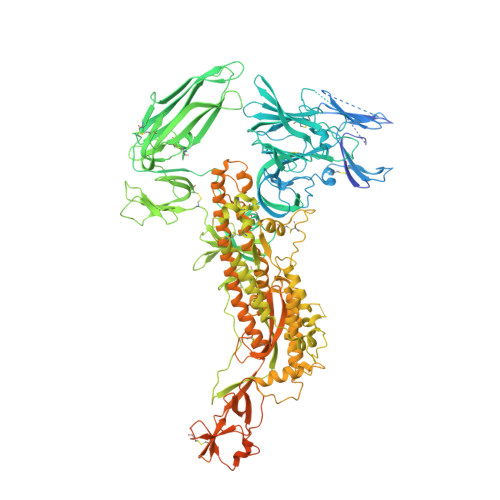
    Porcine epidemic diarrhea virus (PEDV) is an alphacoronavirus that has a significant agricultural and economic impact due to the high mortality rate associated with infection of neonatal piglets. Like other coronaviruses, PEDV makes use of a large, trimeric spike (S) glycoprotein to mediate membrane fusion and gain entry into host cells. Despite the importance of the spike protein in viral entry and host immune responses, high-resolution structural information concerning this large macromolecular machine has been difficult to obtain. Here, we report the cryo-electron microscopy structure of the PEDV S protein in the prefusion conformation at a resolution of 3.1 Å. Our studies revealed that the sialic acid-binding domain at the N terminus of the S1 subunit has an orientation that is substantially different from that observed in the previously determined spike structure from human alphacoronavirus NL63. We also observed dissociated S1 subunit trimers wherein the putative receptor-binding domains exist in a conformation differing from that observed in the intact spike proteins, suggesting that the PEDV receptor-binding domain undergoes conformational rearrangements akin to those that have been described in the related betacoronaviruses. Collectively, these data provide new insights into the biological processes that mediate alphacoronavirus attachment, receptor engagement, and fusion triggering while also identifying a source of conformational heterogeneity that could be manipulated to improve PEDV vaccine antigens. IMPORTANCE Coronavirus spike proteins are large, densely glycosylated macromolecular machines that mediate receptor binding and membrane fusion to facilitate entry into host cells. This report describes the atomic-resolution structure of the spike protein from porcine epidemic diarrhea virus, a pathogenic alphacoronavirus that causes severe agricultural damage. The structure reveals a novel position for the sialic acid-binding attachment domain in the intact spike. We also observed shed fusion-suppressive capping subunits that displayed the putative receptor-binding domain in an accessible conformation. These observations provide a basis for understanding the molecular mechanisms that drive the earliest stages of alphacoronavirus infection and will inform future efforts to rationally design vaccines.


  • Organizational Affiliation

    Department of Molecular Biosciences, The University of Texas at Austin, Austin, Texas, USA.


Macromolecules
Find similar proteins by:  (by identity cutoff)  |  3D Structure
Entity ID: 1
MoleculeChains Sequence LengthOrganismDetailsImage
Spike glycoprotein
A, B, C
1,399Porcine epidemic diarrhea virus CV777Mutation(s): 0 
Gene Names: S2
UniProt
Find proteins for Q91AV1 (Porcine epidemic diarrhea virus (strain CV777))
Explore Q91AV1 
Go to UniProtKB:  Q91AV1
Entity Groups  
Sequence Clusters30% Identity50% Identity70% Identity90% Identity95% Identity100% Identity
UniProt GroupQ91AV1
Sequence Annotations
Expand
  • Reference Sequence
Oligosaccharides

Help

Entity ID: 2
MoleculeChains Length2D Diagram Glycosylation3D Interactions
alpha-D-mannopyranose-(1-2)-alpha-D-mannopyranose-(1-3)-[alpha-D-mannopyranose-(1-6)]beta-D-mannopyranose-(1-4)-2-acetamido-2-deoxy-beta-D-glucopyranose-(1-4)-2-acetamido-2-deoxy-beta-D-glucopyranose
D, I, N
6N-Glycosylation
Glycosylation Resources
GlyTouCan:  G56014GC
GlyCosmos:  G56014GC
GlyGen:  G56014GC
Entity ID: 3
MoleculeChains Length2D Diagram Glycosylation3D Interactions
2-acetamido-2-deoxy-beta-D-glucopyranose-(1-4)-2-acetamido-2-deoxy-beta-D-glucopyranose
E, F, G, H, J
E, F, G, H, J, K, L, M, O, P, Q, R
2N/A
Glycosylation Resources
GlyTouCan:  G42666HT
GlyCosmos:  G42666HT
GlyGen:  G42666HT
Small Molecules
Ligands 1 Unique
IDChains Name / Formula / InChI Key2D Diagram3D Interactions
NAG
Query on NAG

Download Ideal Coordinates CCD File 
AA [auth A]
AB [auth C]
BA [auth A]
BB [auth C]
CA [auth A]
AA [auth A],
AB [auth C],
BA [auth A],
BB [auth C],
CA [auth A],
CB [auth C],
DA [auth A],
DB [auth C],
EA [auth A],
EB [auth C],
FA [auth B],
GA [auth B],
HA [auth B],
IA [auth B],
JA [auth B],
KA [auth B],
LA [auth B],
MA [auth B],
NA [auth B],
OA [auth B],
PA [auth B],
QA [auth B],
RA [auth B],
S [auth A],
SA [auth C],
T [auth A],
TA [auth C],
U [auth A],
UA [auth C],
V [auth A],
VA [auth C],
W [auth A],
WA [auth C],
X [auth A],
XA [auth C],
Y [auth A],
YA [auth C],
Z [auth A],
ZA [auth C]
2-acetamido-2-deoxy-beta-D-glucopyranose
C8 H15 N O6
OVRNDRQMDRJTHS-FMDGEEDCSA-N
Experimental Data & Validation

Experimental Data

  • Method: ELECTRON MICROSCOPY
  • Resolution: 3.14 Å
  • Aggregation State: PARTICLE 
  • Reconstruction Method: SINGLE PARTICLE 
EM Software:
TaskSoftware PackageVersion
RECONSTRUCTIONcryoSPARC2.4.6

Structure Validation

View Full Validation Report


Entry History & Funding Information

Deposition Data


Funding OrganizationLocationGrant Number
National Institutes of Health/National Institute Of Allergy and Infectious Diseases (NIH/NIAID)United StatesR01-AI127521

Revision History  (Full details and data files)

  • Version 1.0: 2019-09-18
    Type: Initial release
  • Version 1.1: 2019-10-02
    Changes: Data collection, Database references
  • Version 1.2: 2019-11-27
    Changes: Database references
  • Version 1.3: 2019-12-18
    Changes: Author supporting evidence
  • Version 2.0: 2020-07-29
    Type: Remediation
    Reason: Carbohydrate remediation
    Changes: Advisory, Atomic model, Data collection, Derived calculations, Structure summary