6U3Y

Structure-based discovery of a novel small-molecule inhibitor of methicillin-resistant S. aureus


Experimental Data Snapshot

  • Method: X-RAY DIFFRACTION
  • Resolution: 2.04 Å
  • R-Value Free: 0.225 
  • R-Value Work: 0.182 
  • R-Value Observed: 0.184 

wwPDB Validation   3D Report Full Report


Ligand Structure Quality Assessment 


This is version 1.3 of the entry. See complete history


Literature

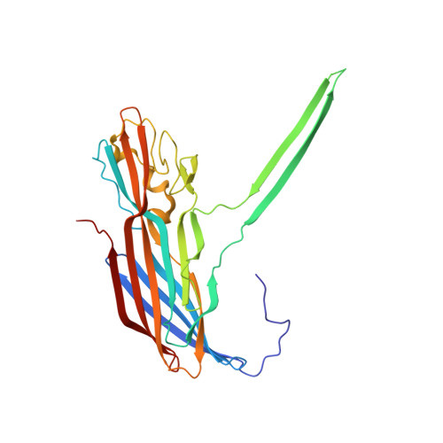
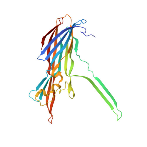
Structure-based discovery of a small-molecule inhibitor of methicillin-resistantStaphylococcus aureusvirulence.

Liu, J.Kozhaya, L.Torres, V.J.Unutmaz, D.Lu, M.

(2020) J Biol Chem 295: 5944-5959

  • DOI: https://doi.org/10.1074/jbc.RA120.012697
  • Primary Citation of Related Structures:  
    6U2S, 6U33, 6U3F, 6U3T, 6U3Y, 6U49, 6U4P

  • PubMed Abstract: 

    The rapid emergence and dissemination of methicillin-resistant Staphylococcus aureus (MRSA) strains poses a major threat to public health. MRSA possesses an arsenal of secreted host-damaging virulence factors that mediate pathogenicity and blunt immune defenses. Panton-Valentine leukocidin (PVL) and α-toxin are exotoxins that create lytic pores in the host cell membrane. They are recognized as being important for the development of invasive MRSA infections and are thus potential targets for antivirulence therapies. Here, we report the high-resolution X-ray crystal structures of both PVL and α-toxin in their soluble, monomeric, and oligomeric membrane-inserted pore states in complex with n -tetradecylphosphocholine (C 14 PC). The structures revealed two evolutionarily conserved phosphatidylcholine-binding mechanisms and their roles in modulating host cell attachment, oligomer assembly, and membrane perforation. Moreover, we demonstrate that the soluble C 14 PC compound protects primary human immune cells in vitro against cytolysis by PVL and α-toxin and hence may serve as the basis for the development of an antivirulence agent for managing MRSA infections.


  • Organizational Affiliation

    Public Health Research Institute, Department of Microbiology, Biochemistry, and Molecular Genetics, New Jersey Medical School, Rutgers University, Newark, New Jersey 07103.


Macromolecules
Find similar proteins by:  (by identity cutoff)  |  3D Structure
Entity ID: 1
MoleculeChains Sequence LengthOrganismDetailsImage
Panton-Valentine Leucocidin F301Staphylococcus aureusMutation(s): 0 
Gene Names: lukF-PVhlgB_1lukF- PVLukPV-FlukPVFNCTC6133_01921
Membrane Entity: Yes 
UniProt
Find proteins for Q5FBD2 (Staphylococcus aureus)
Explore Q5FBD2 
Go to UniProtKB:  Q5FBD2
Entity Groups  
Sequence Clusters30% Identity50% Identity70% Identity90% Identity95% Identity100% Identity
UniProt GroupQ5FBD2
Sequence Annotations
Expand
  • Reference Sequence
Find similar proteins by:  (by identity cutoff)  |  3D Structure
Entity ID: 2
MoleculeChains Sequence LengthOrganismDetailsImage
Gamma-hemolysin subunit AB,
C [auth D]
284Staphylococcus aureusMutation(s): 0 
Gene Names: lukS-PVLukPV-SlukPVSEP54_02590EQ90_09955
UniProt
Find proteins for B1Q017 (Staphylococcus aureus)
Explore B1Q017 
Go to UniProtKB:  B1Q017
Entity Groups  
Sequence Clusters30% Identity50% Identity70% Identity90% Identity95% Identity100% Identity
UniProt GroupB1Q017
Sequence Annotations
Expand
  • Reference Sequence
Small Molecules
Ligands 3 Unique
IDChains Name / Formula / InChI Key2D Diagram3D Interactions
PQJ
Query on PQJ

Download Ideal Coordinates CCD File 
D [auth A],
E [auth A],
F [auth A]
fos-choline-14
C19 H43 N O4 P
JOPCRKJIQSCIFM-UHFFFAOYSA-N
SO4
Query on SO4

Download Ideal Coordinates CCD File 
AA [auth D]
BA [auth D]
CA [auth D]
G [auth A]
H [auth A]
AA [auth D],
BA [auth D],
CA [auth D],
G [auth A],
H [auth A],
I [auth A],
J [auth A],
K [auth A],
L [auth A],
P [auth B],
Q [auth B],
R [auth B],
S [auth B],
X [auth D],
Y [auth D],
Z [auth D]
SULFATE ION
O4 S
QAOWNCQODCNURD-UHFFFAOYSA-L
ACT
Query on ACT

Download Ideal Coordinates CCD File 
DA [auth D]
M [auth A]
N [auth A]
O [auth A]
T [auth B]
DA [auth D],
M [auth A],
N [auth A],
O [auth A],
T [auth B],
U [auth B],
V [auth B],
W [auth B]
ACETATE ION
C2 H3 O2
QTBSBXVTEAMEQO-UHFFFAOYSA-M
Experimental Data & Validation

Experimental Data

  • Method: X-RAY DIFFRACTION
  • Resolution: 2.04 Å
  • R-Value Free: 0.225 
  • R-Value Work: 0.182 
  • R-Value Observed: 0.184 
  • Space Group: I 4 2 2
Unit Cell:
Length ( Å )Angle ( ˚ )
a = 139.097α = 90
b = 139.097β = 90
c = 246.77γ = 90
Software Package:
Software NamePurpose
REFMACrefinement
PDB_EXTRACTdata extraction
HKL-2000data reduction
HKL-2000data scaling
PHASERphasing

Structure Validation

View Full Validation Report


Ligand Structure Quality Assessment 


Entry History & Funding Information

Deposition Data


Funding OrganizationLocationGrant Number
National Institutes of Health/National Human Genome Research Institute (NIH/NHGRI)United States--

Revision History  (Full details and data files)

  • Version 1.0: 2020-03-25
    Type: Initial release
  • Version 1.1: 2020-04-01
    Changes: Database references
  • Version 1.2: 2020-05-13
    Changes: Database references
  • Version 1.3: 2023-10-11
    Changes: Data collection, Database references, Refinement description