6TUW

human XPG-DNA, Complex 1


Experimental Data Snapshot

  • Method: X-RAY DIFFRACTION
  • Resolution: 3.50 Å
  • R-Value Free: 0.293 
  • R-Value Work: 0.265 
  • R-Value Observed: 0.267 

wwPDB Validation   3D Report Full Report


This is version 1.2 of the entry. See complete history


Literature

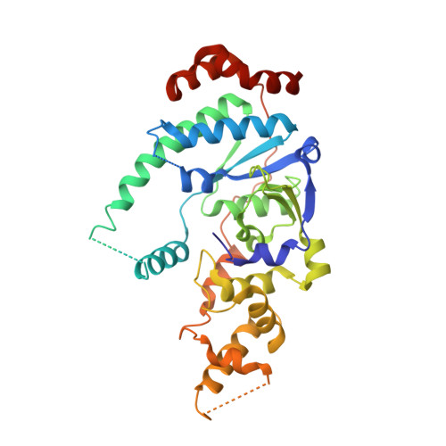
The crystal structure of human XPG, the xeroderma pigmentosum group G endonuclease, provides insight into nucleotide excision DNA repair.

Gonzalez-Corrochano, R.Ruiz, F.M.Taylor, N.M.I.Huecas, S.Drakulic, S.Spinola-Amilibia, M.Fernandez-Tornero, C.

(2020) Nucleic Acids Res 48: 9943-9958

  • DOI: https://doi.org/10.1093/nar/gkaa688
  • Primary Citation of Related Structures:  
    6TUR, 6TUS, 6TUW, 6TUX

  • PubMed Abstract: 

    Nucleotide excision repair (NER) is an essential pathway to remove bulky lesions affecting one strand of DNA. Defects in components of this repair system are at the ground of genetic diseases such as xeroderma pigmentosum (XP) and Cockayne syndrome (CS). The XP complementation group G (XPG) endonuclease cleaves the damaged DNA strand on the 3' side of the lesion coordinated with DNA re-synthesis. Here, we determined crystal structures of the XPG nuclease domain in the absence and presence of DNA. The overall fold exhibits similarities to other flap endonucleases but XPG harbors a dynamic helical arch that is uniquely oriented and defines a gateway. DNA binding through a helix-2-turn-helix motif, assisted by one flanking α-helix on each side, shows high plasticity, which is likely relevant for DNA scanning. A positively-charged canyon defined by the hydrophobic wedge and β-pin motifs provides an additional DNA-binding surface. Mutational analysis identifies helical arch residues that play critical roles in XPG function. A model for XPG participation in NER is proposed. Our structures and biochemical data represent a valuable tool to understand the atomic ground of XP and CS, and constitute a starting point for potential therapeutic applications.


  • Organizational Affiliation

    Centro de Investigaciones Biológicas Margarita Salas, CSIC, Ramiro de Maeztu 9, 28040 Madrid, Spain.


Macromolecules

Find similar proteins by:  (by identity cutoff)  |  3D Structure
Entity ID: 1
MoleculeChains Sequence LengthOrganismDetailsImage
DNA repair protein complementing XP-G cells,DNA repair protein complementing XP-G cells355Homo sapiensMutation(s): 0 
Gene Names: ERCC5ERCM2XPGXPGC
EC: 3.1
UniProt & NIH Common Fund Data Resources
Find proteins for P28715 (Homo sapiens)
Explore P28715 
Go to UniProtKB:  P28715
PHAROS:  P28715
GTEx:  ENSG00000134899 
Entity Groups  
Sequence Clusters30% Identity50% Identity70% Identity90% Identity95% Identity100% Identity
UniProt GroupP28715
Sequence Annotations
Expand
  • Reference Sequence

Find similar nucleic acids by:  Sequence   |   3D Structure  

Entity ID: 2
MoleculeChains LengthOrganismImage
DNA (5'-D(P*TP*GP*CP*AP*GP*AP*GP*TP*TP*C)-3')11unidentified
Sequence Annotations
Expand
  • Reference Sequence

Find similar nucleic acids by:  Sequence   |   3D Structure  

Entity ID: 3
MoleculeChains LengthOrganismImage
DNA (5'-D(P*GP*AP*AP*CP*TP*CP*TP*G)-3')11unidentified
Sequence Annotations
Expand
  • Reference Sequence
Experimental Data & Validation

Experimental Data

  • Method: X-RAY DIFFRACTION
  • Resolution: 3.50 Å
  • R-Value Free: 0.293 
  • R-Value Work: 0.265 
  • R-Value Observed: 0.267 
  • Space Group: I 4 2 2
Unit Cell:
Length ( Å )Angle ( ˚ )
a = 129.51α = 90
b = 129.51β = 90
c = 118.73γ = 90
Software Package:
Software NamePurpose
PHENIXrefinement
XDSdata reduction
XSCALEdata scaling
PHASERphasing

Structure Validation

View Full Validation Report


Entry History 

Deposition Data

Revision History  (Full details and data files)

  • Version 1.0: 2020-09-16
    Type: Initial release
  • Version 1.1: 2020-10-07
    Changes: Database references
  • Version 1.2: 2024-01-24
    Changes: Data collection, Database references, Refinement description