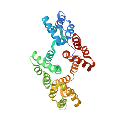
6TU2

Crystal structure of rat annexin A11


Experimental Data Snapshot

  • Method: X-RAY DIFFRACTION
  • Resolution: 2.30 Å
  • R-Value Free: 0.266 
  • R-Value Work: 0.228 
  • R-Value Observed: 0.230 

Starting Model: experimental
View more details

wwPDB Validation   3D Report Full Report


This is version 1.2 of the entry. See complete history


Literature

Structure of the ALS Mutation Target Annexin A11 Reveals a Stabilising N-Terminal Segment.

Lillebostad, P.A.G.Raasakka, A.Hjellbrekke, S.J.Patil, S.Rostbo, T.Hollas, H.Sakya, S.A.Szigetvari, P.D.Vedeler, A.Kursula, P.

(2020) Biomolecules 10

  • DOI: https://doi.org/10.3390/biom10040660
  • Primary Citation of Related Structures:  
    6TU2

  • PubMed Abstract: 

    The functions of the annexin family of proteins involve binding to Ca 2+ , lipid membranes, other proteins, and RNA, and the annexins share a common folded core structure at the C terminus. Annexin A11 (AnxA11) has a long N-terminal region, which is predicted to be disordered, binds RNA, and forms membraneless organelles involved in neuronal transport. Mutations in AnxA11 have been linked to amyotrophic lateral sclerosis (ALS). We studied the structure and stability of AnxA11 and identified a short stabilising segment in the N-terminal end of the folded core, which links domains I and IV. The crystal structure of the AnxA11 core highlights main-chain hydrogen bonding interactions formed through this bridging segment, which are likely conserved in most annexins. The structure was also used to study the currently known ALS mutations in AnxA11. Three of these mutations correspond to buried Arg residues highly conserved in the annexin family, indicating central roles in annexin folding. The structural data provide starting points for detailed structure-function studies of both full-length AnxA11 and the disease variants being identified in ALS.


  • Organizational Affiliation

    Department of Biomedicine, University of Bergen, Jonas Lies vei 91, 5009 Bergen, Norway.


Macromolecules
Find similar proteins by:  (by identity cutoff)  |  3D Structure
Entity ID: 1
MoleculeChains Sequence LengthOrganismDetailsImage
Annexin
A, B, C
326Rattus norvegicusMutation(s): 0 
Gene Names: Anxa11rCG_39189
UniProt
Find proteins for Q5XI77 (Rattus norvegicus)
Explore Q5XI77 
Go to UniProtKB:  Q5XI77
Entity Groups  
Sequence Clusters30% Identity50% Identity70% Identity90% Identity95% Identity100% Identity
UniProt GroupQ5XI77
Sequence Annotations
Expand
  • Reference Sequence
Small Molecules
Ligands 1 Unique
IDChains Name / Formula / InChI Key2D Diagram3D Interactions
CA (Subject of Investigation/LOI)
Query on CA

Download Ideal Coordinates CCD File 
D [auth A]
E [auth A]
F [auth A]
G [auth B]
H [auth B]
D [auth A],
E [auth A],
F [auth A],
G [auth B],
H [auth B],
I [auth B],
J [auth B],
K [auth C],
L [auth C],
M [auth C]
CALCIUM ION
Ca
BHPQYMZQTOCNFJ-UHFFFAOYSA-N
Experimental Data & Validation

Experimental Data

  • Method: X-RAY DIFFRACTION
  • Resolution: 2.30 Å
  • R-Value Free: 0.266 
  • R-Value Work: 0.228 
  • R-Value Observed: 0.230 
  • Space Group: P 1
Unit Cell:
Length ( Å )Angle ( ˚ )
a = 39.07α = 114
b = 86.69β = 101.98
c = 87.3γ = 97.21
Software Package:
Software NamePurpose
PHENIXrefinement
XDSdata reduction
XDSdata scaling
PHASERphasing

Structure Validation

View Full Validation Report


Entry History 

Deposition Data

Revision History  (Full details and data files)

  • Version 1.0: 2020-05-06
    Type: Initial release
  • Version 1.1: 2020-11-18
    Changes: Database references, Derived calculations
  • Version 1.2: 2024-01-24
    Changes: Data collection, Database references, Refinement description