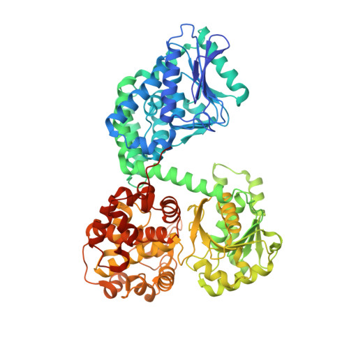
6TNM

E. coli aerobic trifunctional enzyme subunit-alpha


Experimental Data Snapshot

  • Method: X-RAY DIFFRACTION
  • Resolution: 2.95 Å
  • R-Value Free: 0.271 
  • R-Value Work: 0.230 
  • R-Value Observed: 0.232 

Starting Model: experimental
View more details

wwPDB Validation   3D Report Full Report


Ligand Structure Quality Assessment 


This is version 1.2 of the entry. See complete history


Literature

Insights into the stability and substrate specificity of the E. coli aerobic beta-oxidation trifunctional enzyme complex.

Sah-Teli, S.K.Hynonen, M.J.Sulu, R.Dalwani, S.Schmitz, W.Wierenga, R.K.Venkatesan, R.

(2020) J Struct Biol 210: 107494-107494

  • DOI: https://doi.org/10.1016/j.jsb.2020.107494
  • Primary Citation of Related Structures:  
    6TNM

  • PubMed Abstract: 

    Degradation of fatty acids by the β-oxidation pathway results in the formation of acetyl-CoA which enters the TCA cycle for the production of ATP. In E. coli, the last three steps of the β-oxidation are catalyzed by two heterotetrameric α 2 β 2 enzymes namely the aerobic trifunctional enzyme (EcTFE) and the anaerobic TFE (anEcTFE). The α-subunit of TFE has 2E-enoyl-CoA hydratase (ECH) and 3S-hydroxyacyl-CoA dehydrogenase (HAD) activities whereas the β-subunit is a thiolase with 3-ketoacyl-CoA thiolase (KAT) activity. Recently, it has been shown that the two TFEs have complementary substrate specificities allowing for the complete degradation of long chain fatty acyl-CoAs into acetyl-CoA under aerobic conditions. Also, it has been shown that the tetrameric EcTFE and anEcTFE assemblies are similar to the TFEs of Pseudomans fragi and human, respectively. Here the properties of the EcTFE subunits are further characterized. Strikingly, it is observed that when expressed separately, EcTFE-α is a catalytically active monomer whereas EcTFE-β is inactive. However, when mixed together active EcTFE tetramer is reconstituted. The crystal structure of the EcTFE-α chain is also reported, complexed with ATP, bound in its HAD active site. Structural comparisons show that the EcTFE hydratase active site has a relatively small fatty acyl tail binding pocket when compared to other TFEs in good agreement with its preferred specificity for short chain 2E-enoyl-CoA substrates. Furthermore, it is observed that millimolar concentrations of ATP destabilize the EcTFE complex, and this may have implications for the ATP-mediated regulation of β-oxidation in E. coli.


  • Organizational Affiliation

    Faculty of Biochemistry and Molecular Medicine, and Biocenter Oulu, University of Oulu, Oulu, Finland.


Macromolecules
Find similar proteins by:  (by identity cutoff)  |  3D Structure
Entity ID: 1
MoleculeChains Sequence LengthOrganismDetailsImage
Fatty acid oxidation complex subunit alpha743Escherichia coli K-12Mutation(s): 0 
Gene Names: fadBoldBb3846JW3822
EC: 4.2.1.17 (PDB Primary Data), 5.1.2.3 (PDB Primary Data), 5.3.3.8 (PDB Primary Data), 1.1.1.35 (PDB Primary Data)
UniProt
Find proteins for P21177 (Escherichia coli (strain K12))
Explore P21177 
Go to UniProtKB:  P21177
Entity Groups  
Sequence Clusters30% Identity50% Identity70% Identity90% Identity95% Identity100% Identity
UniProt GroupP21177
Sequence Annotations
Expand
  • Reference Sequence
Small Molecules
Ligands 3 Unique
IDChains Name / Formula / InChI Key2D Diagram3D Interactions
ATP (Subject of Investigation/LOI)
Query on ATP

Download Ideal Coordinates CCD File 
C [auth A]ADENOSINE-5'-TRIPHOSPHATE
C10 H16 N5 O13 P3
ZKHQWZAMYRWXGA-KQYNXXCUSA-N
GOL
Query on GOL

Download Ideal Coordinates CCD File 
B [auth A]GLYCEROL
C3 H8 O3
PEDCQBHIVMGVHV-UHFFFAOYSA-N
NO3
Query on NO3

Download Ideal Coordinates CCD File 
D [auth A]
E [auth A]
F [auth A]
G [auth A]
H [auth A]
D [auth A],
E [auth A],
F [auth A],
G [auth A],
H [auth A],
I [auth A],
J [auth A]
NITRATE ION
N O3
NHNBFGGVMKEFGY-UHFFFAOYSA-N
Experimental Data & Validation

Experimental Data

  • Method: X-RAY DIFFRACTION
  • Resolution: 2.95 Å
  • R-Value Free: 0.271 
  • R-Value Work: 0.230 
  • R-Value Observed: 0.232 
  • Space Group: C 2 2 21
Unit Cell:
Length ( Å )Angle ( ˚ )
a = 130.384α = 90
b = 214.113β = 90
c = 76.52γ = 90
Software Package:
Software NamePurpose
xia2data reduction
DIALSdata scaling
MoRDaphasing
PHENIXrefinement
Cootmodel building

Structure Validation

View Full Validation Report


Ligand Structure Quality Assessment 


Entry History & Funding Information

Deposition Data


Funding OrganizationLocationGrant Number
Academy of FinlandFinland289024, 293369, 319194

Revision History  (Full details and data files)

  • Version 1.0: 2020-03-25
    Type: Initial release
  • Version 1.1: 2020-05-20
    Changes: Database references
  • Version 1.2: 2024-01-24
    Changes: Data collection, Database references, Refinement description