6TIS | pdb_00006tis

DROSOPHILA GDP-TUBULIN


Experimental Data Snapshot

  • Method: X-RAY DIFFRACTION
  • Resolution: 2.30 Å
  • R-Value Free: 
    0.218 (Depositor), 0.236 (DCC) 
  • R-Value Work: 
    0.183 (Depositor), 0.197 (DCC) 
  • R-Value Observed: 
    0.185 (Depositor) 

Starting Model: experimental
View more details

wwPDB Validation 3D Report Full Report

Validation slider image for 6TIS

Ligand Structure Quality Assessment 


This is version 1.2 of the entry. See complete history

Literature

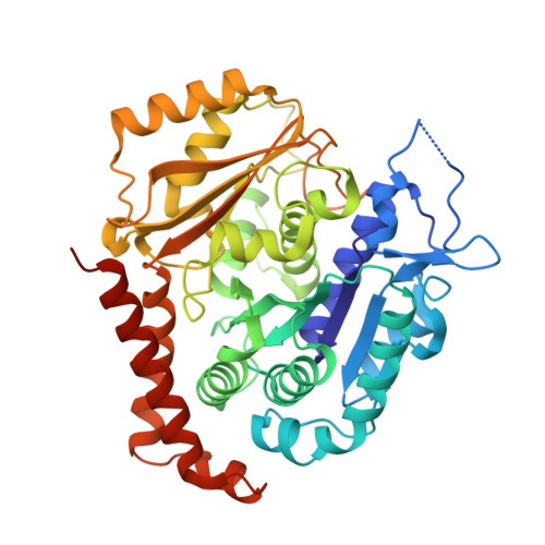
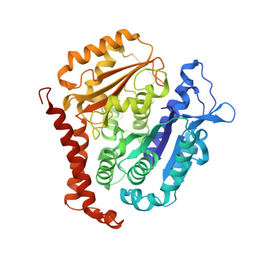
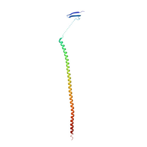
GTP-dependent formation of straight tubulin oligomers leads to microtubule nucleation.

Ayukawa, R.Iwata, S.Imai, H.Kamimura, S.Hayashi, M.Ngo, K.X.Minoura, I.Uchimura, S.Makino, T.Shirouzu, M.Shigematsu, H.Sekimoto, K.Gigant, B.Muto, E.

(2021) J Cell Biol 220

  • DOI: https://doi.org/10.1083/jcb.202007033
  • Primary Citation Related Structures: 
    6TIS, 6TIU, 6TIY, 6TIZ

  • PubMed Abstract: 

    Nucleation of microtubules (MTs) is essential for cellular activities, but its mechanism is unknown because of the difficulty involved in capturing rare stochastic events in the early stage of polymerization. Here, combining rapid flush negative stain electron microscopy (EM) and kinetic analysis, we demonstrate that the formation of straight oligomers of critical size is essential for nucleation. Both GDP and GTP tubulin form single-stranded oligomers with a broad range of curvatures, but upon nucleation, the curvature distribution of GTP oligomers is shifted to produce a minor population of straight oligomers. With tubulin having the Y222F mutation in the β subunit, the proportion of straight oligomers increases and nucleation accelerates. Our results support a model in which GTP binding generates a minor population of straight oligomers compatible with lateral association and further growth to MTs. This study suggests that cellular factors involved in nucleation promote it via stabilization of straight oligomers.


  • Organizational Affiliation
    • Laboratory for Molecular Biophysics, RIKEN Center for Brain Science, Saitama, Japan.

Macromolecule Content 

  • Total Structure Weight: 220.44 kDa 
  • Atom Count: 15,255 
  • Modeled Residue Count: 1,850 
  • Deposited Residue Count: 1,937 
  • Unique protein chains: 3

Macromolecules

Find similar proteins by:|  3D Structure
Entity ID: 1
MoleculeChains  Sequence LengthOrganismDetailsImage
Tubulin alpha-1 chain
A, C
450Drosophila melanogasterMutation(s): 1 
Gene Names: alphaTub84BtubA84BCG1913
EC: 3.6.5
UniProt
Find proteins for P06603 (Drosophila melanogaster)
Explore P06603 
Go to UniProtKB:  P06603
Entity Groups
Sequence Clusters30% Identity50% Identity70% Identity90% Identity95% Identity100% Identity
UniProt GroupP06603
Sequence Annotations
Expand
Reference Sequence
Find similar proteins by:|  3D Structure
Entity ID: 2
MoleculeChains  Sequence LengthOrganismDetailsImage
Tubulin beta-1 chain
B, D
447Drosophila melanogasterMutation(s): 0 
Gene Names: betaTub56DCG9277
UniProt
Find proteins for Q24560 (Drosophila melanogaster)
Explore Q24560 
Go to UniProtKB:  Q24560
Entity Groups
Sequence Clusters30% Identity50% Identity70% Identity90% Identity95% Identity100% Identity
UniProt GroupQ24560
Sequence Annotations
Expand
Reference Sequence
Find similar proteins by:|  3D Structure
Entity ID: 3
MoleculeChains  Sequence LengthOrganismDetailsImage
Stathmin-4143Rattus norvegicusMutation(s): 3 
Gene Names: Stmn4
UniProt
Find proteins for P63043 (Rattus norvegicus)
Explore P63043 
Go to UniProtKB:  P63043
Entity Groups
Sequence Clusters30% Identity50% Identity70% Identity90% Identity95% Identity100% Identity
UniProt GroupP63043
Sequence Annotations
Expand
Reference Sequence

Small Molecules

Ligands 5 Unique
IDChains Name / Formula / InChI Key2D Diagram3D Interactions
GTP

Query on GTP


Download:Ideal Coordinates CCD File
F [auth A],
R [auth C]
GUANOSINE-5'-TRIPHOSPHATE
C10 H16 N5 O14 P3
XKMLYUALXHKNFT-UUOKFMHZSA-N
GDP

Query on GDP


Download:Ideal Coordinates CCD File
M [auth B],
U [auth D]
GUANOSINE-5'-DIPHOSPHATE
C10 H15 N5 O11 P2
QGWNDRXFNXRZMB-UUOKFMHZSA-N
SO4

Query on SO4


Download:Ideal Coordinates CCD File
H [auth A]
I [auth A]
J [auth A]
K [auth A]
N [auth B]
H [auth A],
I [auth A],
J [auth A],
K [auth A],
N [auth B],
O [auth B],
P [auth B],
T [auth C],
V [auth D],
W [auth D],
X [auth D]
SULFATE ION
O4 S
QAOWNCQODCNURD-UHFFFAOYSA-L
GOL

Query on GOL


Download:Ideal Coordinates CCD File
L [auth A],
Q [auth B]
GLYCEROL
C3 H8 O3
PEDCQBHIVMGVHV-UHFFFAOYSA-N
MG

Query on MG


Download:Ideal Coordinates CCD File
G [auth A],
S [auth C]
MAGNESIUM ION
Mg
JLVVSXFLKOJNIY-UHFFFAOYSA-N

Experimental Data & Validation

Experimental Data

  • Method: X-RAY DIFFRACTION
  • Resolution: 2.30 Å
  • R-Value Free:  0.218 (Depositor), 0.236 (DCC) 
  • R-Value Work:  0.183 (Depositor), 0.197 (DCC) 
  • R-Value Observed: 0.185 (Depositor) 
Space Group: P 21 21 21
Unit Cell:
Length ( Å )Angle ( ˚ )
a = 66.588α = 90
b = 126.515β = 90
c = 250.241γ = 90
Software Package:
Software NamePurpose
BUSTERrefinement
XDSdata reduction
Aimlessdata scaling
BUSTERphasing

Structure Validation

View Full Validation Report


Ligand Structure Quality Assessment 


Entry History 

Deposition Data

  • Released Date: 2021-01-27 
  • Deposition Author(s): Gigant, B.

Revision History  (Full details and data files)

  • Version 1.0: 2021-01-27
    Type: Initial release
  • Version 1.1: 2021-02-17
    Changes: Database references
  • Version 1.2: 2024-01-24
    Changes: Data collection, Database references, Refinement description