6TDE | pdb_00006tde

Tubulin-inhibitor complex


Experimental Data Snapshot

  • Method: X-RAY DIFFRACTION
  • Resolution: 2.29 Å
  • R-Value Free: 
    0.232 (Depositor), 0.251 (DCC) 
  • R-Value Work: 
    0.198 (Depositor) 
  • R-Value Observed: 
    0.199 (Depositor) 

Starting Model: experimental
View more details

wwPDB Validation 3D Report Full Report

Validation slider image for 6TDE

Ligand Structure Quality Assessment 


This is version 1.2 of the entry. See complete history

Literature

Discovery of dihydrofuranoallocolchicinoids - Highly potent antimitotic agents with low acute toxicity.

Shchegravina, E.S.Svirshchevskaya, E.V.Combes, S.Allegro, D.Barbier, P.Gigant, B.Varela, P.F.Gavryushin, A.E.Kobanova, D.A.Shchekotikhin, A.E.Fedorov, A.Y.

(2020) Eur J Med Chem 207: 112724-112724

  • DOI: https://doi.org/10.1016/j.ejmech.2020.112724
  • Primary Citation Related Structures: 
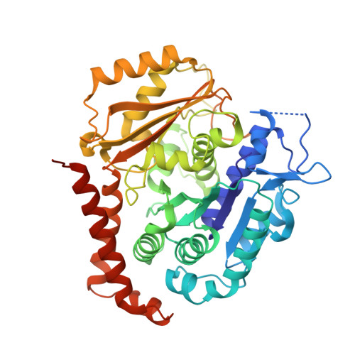
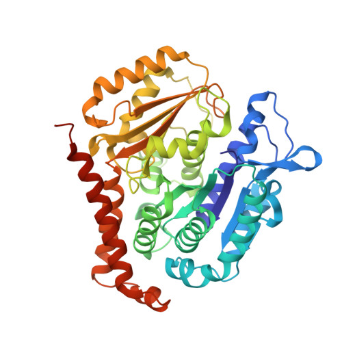
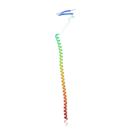
    6TDE

  • PubMed Abstract: 

    Two series of heterocyclic colchicinoids bearing β-methylenedihydrofuran or 2H-pyran-2-one fragments were synthesized by the intramolecular Heck reaction. Methylenedihydrofuran compounds 9a and 9h were found to be the most cytotoxic among currently known colchicinoids, exhibiting outstanding antiproliferative activity on tumor cell lines in picomolar (0.01-2.1 nM) range of concentrations. Compound 9a potently and substoichiometrically inhibits microtubule formation in vitro, being an order of magnitude more active in this assay than colchicine. Derivatives 9a and 9h revealed relatively low acute toxicity in mice (LD 50  ≥ 10 mg/kg i.v.). The X-Ray structure of colchicinoid 9a bound to tubulin confirmed interaction of this compound with the colchicine binding site of tubulin.


  • Organizational Affiliation
    • Department of Organic Chemistry, Nizhny Novgorod State University, Gagarina Av. 23, Nizhny Novgorod, 603950, Russian Federation.

Macromolecule Content 

  • Total Structure Weight: 220.79 kDa 
  • Atom Count: 15,022 
  • Modeled Residue Count: 1,858 
  • Deposited Residue Count: 1,935 
  • Unique protein chains: 3

Macromolecules

Find similar proteins by:|  3D Structure
Entity ID: 1
MoleculeChains  Sequence LengthOrganismDetailsImage
Tubulin alpha chain
A, C
451Ovis ariesMutation(s): 0 
UniProt
Find proteins for D0VWZ0 (Ovis aries)
Explore D0VWZ0 
Go to UniProtKB:  D0VWZ0
Entity Groups
Sequence Clusters30% Identity50% Identity70% Identity90% Identity95% Identity100% Identity
UniProt GroupD0VWZ0
Sequence Annotations
Expand
Reference Sequence
Find similar proteins by:|  3D Structure
Entity ID: 2
MoleculeChains  Sequence LengthOrganismDetailsImage
Tubulin beta chain
B, D
445Ovis ariesMutation(s): 0 
UniProt
Find proteins for D0VWY9 (Ovis aries)
Explore D0VWY9 
Go to UniProtKB:  D0VWY9
Entity Groups
Sequence Clusters30% Identity50% Identity70% Identity90% Identity95% Identity100% Identity
UniProt GroupD0VWY9
Sequence Annotations
Expand
Reference Sequence
Find similar proteins by:|  3D Structure
Entity ID: 3
MoleculeChains  Sequence LengthOrganismDetailsImage
Stathmin-4143Rattus norvegicusMutation(s): 3 
Gene Names: Stmn4
UniProt
Find proteins for P63043 (Rattus norvegicus)
Explore P63043 
Go to UniProtKB:  P63043
Entity Groups
Sequence Clusters30% Identity50% Identity70% Identity90% Identity95% Identity100% Identity
UniProt GroupP63043
Sequence Annotations
Expand
Reference Sequence

Small Molecules

Ligands 5 Unique
IDChains Name / Formula / InChI Key2D Diagram3D Interactions
GTP

Query on GTP


Download:Ideal Coordinates CCD File
G [auth A],
P [auth C]
GUANOSINE-5'-TRIPHOSPHATE
C10 H16 N5 O14 P3
XKMLYUALXHKNFT-UUOKFMHZSA-N
GDP

Query on GDP


Download:Ideal Coordinates CCD File
K [auth B],
U [auth D]
GUANOSINE-5'-DIPHOSPHATE
C10 H15 N5 O11 P2
QGWNDRXFNXRZMB-UUOKFMHZSA-N
N3Z
(Subject of Investigation/LOI)

Query on N3Z


Download:Ideal Coordinates CCD File
L [auth B],
V [auth D]
~{N}-[(10~{S})-3,4,5-trimethoxy-16-methylidene-14-oxatetracyclo[9.7.0.0^{2,7}.0^{13,17}]octadeca-1(18),2,4,6,11,13(17)-hexaen-10-yl]ethanamide
C23 H25 N O5
DQVCQNGCZCOGHU-SFHVURJKSA-N
SO4

Query on SO4


Download:Ideal Coordinates CCD File
F [auth A]
I [auth B]
J [auth B]
M [auth C]
N [auth C]
F [auth A],
I [auth B],
J [auth B],
M [auth C],
N [auth C],
O [auth C],
R [auth D],
S [auth D],
T [auth D]
SULFATE ION
O4 S
QAOWNCQODCNURD-UHFFFAOYSA-L
MG

Query on MG


Download:Ideal Coordinates CCD File
H [auth A],
Q [auth C]
MAGNESIUM ION
Mg
JLVVSXFLKOJNIY-UHFFFAOYSA-N

Experimental Data & Validation

Experimental Data

  • Method: X-RAY DIFFRACTION
  • Resolution: 2.29 Å
  • R-Value Free:  0.232 (Depositor), 0.251 (DCC) 
  • R-Value Work:  0.198 (Depositor) 
  • R-Value Observed: 0.199 (Depositor) 
Space Group: P 21 21 21
Unit Cell:
Length ( Å )Angle ( ˚ )
a = 64.477α = 90
b = 128.142β = 90
c = 251.117γ = 90
Software Package:
Software NamePurpose
BUSTERrefinement
XDSdata reduction
autoPROCdata scaling
STARANISOdata scaling
PHASERphasing
Cootmodel building

Structure Validation

View Full Validation Report


Ligand Structure Quality Assessment 


Entry History 

Deposition Data

Revision History  (Full details and data files)

  • Version 1.0: 2020-09-02
    Type: Initial release
  • Version 1.1: 2024-01-24
    Changes: Data collection, Database references, Refinement description
  • Version 1.2: 2024-11-20
    Changes: Structure summary