6TBX

Trypanosoma brucei PTR1 (TbPTR1) in complex with a tricyclic-based inhibitor


Experimental Data Snapshot

  • Method: X-RAY DIFFRACTION
  • Resolution: 1.30 Å
  • R-Value Free: 0.159 
  • R-Value Work: 0.126 
  • R-Value Observed: 0.128 

wwPDB Validation   3D Report Full Report


Ligand Structure Quality Assessment 


This is version 1.2 of the entry. See complete history


Literature

High-resolution crystal structure of Trypanosoma brucei pteridine reductase 1 in complex with an innovative tricyclic-based inhibitor.

Landi, G.Linciano, P.Tassone, G.Costi, M.P.Mangani, S.Pozzi, C.

(2020) Acta Crystallogr D Struct Biol 76: 558-564

  • DOI: https://doi.org/10.1107/S2059798320004891
  • Primary Citation of Related Structures:  
    6TBX

  • PubMed Abstract: 

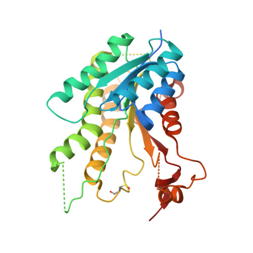
    The protozoan parasite Trypanosoma brucei is the etiological agent of human African trypanosomiasis (HAT). HAT, together with other neglected tropical diseases, causes serious health and economic issues, especially in tropical and subtropical areas. The classical antifolates targeting dihydrofolate reductase (DHFR) are ineffective towards trypanosomatid parasites owing to a metabolic bypass by the expression of pteridine reductase 1 (PTR1). The combined inhibition of PTR1 and DHFR activities in Trypanosoma parasites represents a promising strategy for the development of new effective treatments for HAT. To date, only monocyclic and bicyclic aromatic systems have been proposed as inhibitors of T. brucei PTR1 (TbPTR1); nevertheless, the size of the catalytic cavity allows the accommodation of expanded molecular cores. Here, an innovative tricyclic-based compound has been explored as a TbPTR1-targeting molecule and its potential application for the development of a new class of PTR1 inhibitors has been evaluated. 2,4-Diaminopyrimido[4,5-b]indol-6-ol (1) was designed and synthesized, and was found to be effective in blocking TbPTR1 activity, with a K i in the low-micromolar range. The binding mode of 1 was clarified through the structural characterization of its ternary complex with TbPTR1 and the cofactor NADP(H), which was determined to 1.30 Å resolution. The compound adopts a substrate-like orientation inside the cavity that maximizes the binding contributions of hydrophobic and hydrogen-bond interactions. The binding mode of 1 was compared with those of previously reported bicyclic inhibitors, providing new insights for the design of innovative tricyclic-based molecules targeting TbPTR1.


  • Organizational Affiliation

    Department of Biotechnology, Chemistry and Pharmacy - Department of Excellence 2018-2022, University of Siena, Via Aldo Moro 2, 53100 Siena, Italy.


Macromolecules
Find similar proteins by:  (by identity cutoff)  |  3D Structure
Entity ID: 1
MoleculeChains Sequence LengthOrganismDetailsImage
Pteridine reductase
A, B, D
288Trypanosoma brucei bruceiMutation(s): 0 
Gene Names: PTR1
UniProt
Find proteins for O76290 (Trypanosoma brucei brucei)
Explore O76290 
Go to UniProtKB:  O76290
Entity Groups  
Sequence Clusters30% Identity50% Identity70% Identity90% Identity95% Identity100% Identity
UniProt GroupO76290
Sequence Annotations
Expand
  • Reference Sequence
Find similar proteins by:  (by identity cutoff)  |  3D Structure
Entity ID: 2
MoleculeChains Sequence LengthOrganismDetailsImage
Pteridine reductase288Trypanosoma brucei bruceiMutation(s): 0 
Gene Names: PTR1
UniProt
Find proteins for O76290 (Trypanosoma brucei brucei)
Explore O76290 
Go to UniProtKB:  O76290
Entity Groups  
Sequence Clusters30% Identity50% Identity70% Identity90% Identity95% Identity100% Identity
UniProt GroupO76290
Sequence Annotations
Expand
  • Reference Sequence
Small Molecules
Ligands 4 Unique
IDChains Name / Formula / InChI Key2D Diagram3D Interactions
NAP
Query on NAP

Download Ideal Coordinates CCD File 
E [auth A],
G [auth B],
J [auth C],
M [auth D]
NADP NICOTINAMIDE-ADENINE-DINUCLEOTIDE PHOSPHATE
C21 H28 N7 O17 P3
XJLXINKUBYWONI-NNYOXOHSSA-N
N0Z (Subject of Investigation/LOI)
Query on N0Z

Download Ideal Coordinates CCD File 
F [auth A],
H [auth B],
L [auth C],
N [auth D]
2,4-bis(azanyl)-9~{H}-pyrimido[4,5-b]indol-6-ol
C10 H9 N5 O
QPHUIBRHCXZODB-UHFFFAOYSA-N
GOL
Query on GOL

Download Ideal Coordinates CCD File 
K [auth C]GLYCEROL
C3 H8 O3
PEDCQBHIVMGVHV-UHFFFAOYSA-N
ACT
Query on ACT

Download Ideal Coordinates CCD File 
I [auth B],
O [auth D]
ACETATE ION
C2 H3 O2
QTBSBXVTEAMEQO-UHFFFAOYSA-M
Modified Residues  1 Unique
IDChains TypeFormula2D DiagramParent
CSX
Query on CSX
C
L-PEPTIDE LINKINGC3 H7 N O3 SCYS
Binding Affinity Annotations 
IDSourceBinding Affinity
N0Z Binding MOAD:  6TBX Ki: 8.30e+4 (nM) from 1 assay(s)
Experimental Data & Validation

Experimental Data

  • Method: X-RAY DIFFRACTION
  • Resolution: 1.30 Å
  • R-Value Free: 0.159 
  • R-Value Work: 0.126 
  • R-Value Observed: 0.128 
  • Space Group: P 1 21 1
Unit Cell:
Length ( Å )Angle ( ˚ )
a = 74.677α = 90
b = 90.779β = 115.65
c = 82.81γ = 90
Software Package:
Software NamePurpose
REFMACrefinement
PDB_EXTRACTdata extraction
iMOSFLMdata reduction
SCALAdata scaling
MOLREPphasing

Structure Validation

View Full Validation Report


Ligand Structure Quality Assessment 


Entry History & Funding Information

Deposition Data


Funding OrganizationLocationGrant Number
European Union603240

Revision History  (Full details and data files)

  • Version 1.0: 2020-06-03
    Type: Initial release
  • Version 1.1: 2020-06-17
    Changes: Database references
  • Version 1.2: 2024-01-24
    Changes: Data collection, Database references, Refinement description