Molecular basis of the interaction of the human tyrosine phosphatase PTPN3 with the hepatitis B virus core protein.
Genera, M., Quioc-Salomon, B., Nourisson, A., Colcombet-Cazenave, B., Haouz, A., Mechaly, A., Matondo, M., Duchateau, M., Konig, A., Windisch, M.P., Neuveut, C., Wolff, N., Caillet-Saguy, C.(2021) Sci Rep 11: 944-944
- PubMed: 33441627 Search on PubMedSearch on PubMed Central
- DOI: https://doi.org/10.1038/s41598-020-79580-9
- Primary Citation Related Structures:
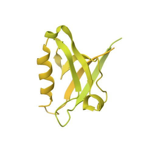
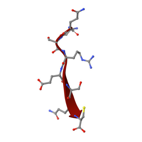
6T36 - PubMed Abstract:
Interactions between the hepatitis B virus core protein (HBc) and host cell proteins are poorly understood, although they may be essential for the propagation of the virus and its pathogenicity. HBc has a C-terminal PDZ (PSD-95, Dlg1, ZO-1)-binding motif (PBM) that is responsible for interactions with host PDZ domain-containing proteins. In this work, we focused on the human protein tyrosine phosphatase non-receptor type 3 (PTPN3) and its interaction with HBc. We solved the crystal structure of the PDZ domain of PTPN3 in complex with the PBM of HBc, revealing a network of interactions specific to class I PDZ domains despite the presence of a C-terminal cysteine in this atypical PBM. We further showed that PTPN3 binds the HBc protein within capsids or as a homodimer. We demonstrate that overexpression of PTPN3 significantly affects HBV infection in HepG2 NTCP cells. Finally, we performed proteomics studies on both sides by pull-down assays and screening of a human PDZ domain library. We identified a pool of human PBM-containing proteins that might interact with PTPN3 in cells and that could be in competition with the HBc PBM during infection, and we also identified potential cellular partners of HBc through PDZ-PBM interactions. This study opens up many avenues of future investigations into the pathophysiology of HBV.
- Channel-Receptors Unit, UMR 3571, CNRS, Institut Pasteur, 75015, Paris, France.
Organizational Affiliation: