6SXB

XPF-ERCC1 Cryo-EM Structure, DNA-Bound form


Experimental Data Snapshot

  • Method: ELECTRON MICROSCOPY
  • Resolution: 7.90 Å
  • Aggregation State: 2D ARRAY 
  • Reconstruction Method: SINGLE PARTICLE 

wwPDB Validation   3D Report Full Report


This is version 1.0 of the entry. See complete history


Literature

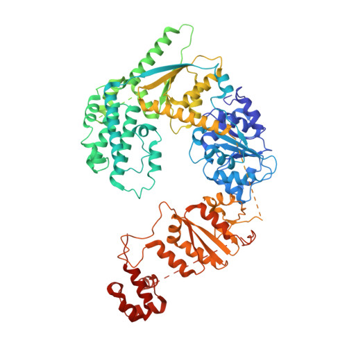
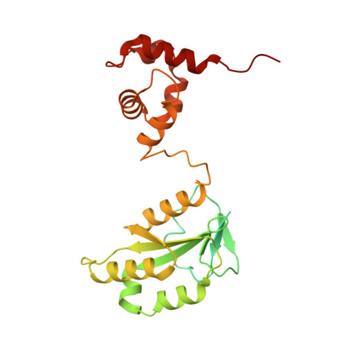
Cryo-EM structures of the XPF-ERCC1 endonuclease reveal how DNA-junction engagement disrupts an auto-inhibited conformation.

Jones, M.Beuron, F.Borg, A.Nans, A.Earl, C.P.Briggs, D.C.Snijders, A.P.Bowles, M.Morris, E.P.Linch, M.McDonald, N.Q.

(2020) Nat Commun 11: 1120-1120

  • DOI: https://doi.org/10.1038/s41467-020-14856-2
  • Primary Citation of Related Structures:  
    6SXA, 6SXB

  • PubMed Abstract: 

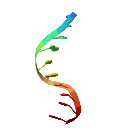
    The structure-specific endonuclease XPF-ERCC1 participates in multiple DNA damage repair pathways including nucleotide excision repair (NER) and inter-strand crosslink repair (ICLR). How XPF-ERCC1 is catalytically activated by DNA junction substrates is not currently understood. Here we report cryo-electron microscopy structures of both DNA-free and DNA-bound human XPF-ERCC1. DNA-free XPF-ERCC1 adopts an auto-inhibited conformation in which the XPF helical domain masks the ERCC1 (HhH) 2 domain and restricts access to the XPF catalytic site. DNA junction engagement releases the ERCC1 (HhH) 2 domain to couple with the XPF-ERCC1 nuclease/nuclease-like domains. Structure-function data indicate xeroderma pigmentosum patient mutations frequently compromise the structural integrity of XPF-ERCC1. Fanconi anaemia patient mutations in XPF often display substantial in-vitro activity but are resistant to activation by ICLR recruitment factor SLX4. Our data provide insights into XPF-ERCC1 architecture and catalytic activation.


  • Organizational Affiliation

    Signalling and Structural Biology Laboratory, Francis Crick Institute, NW1 1AT, London, UK.


Macromolecules

Find similar proteins by:  (by identity cutoff)  |  3D Structure
Entity ID: 1
MoleculeChains Sequence LengthOrganismDetailsImage
DNA repair endonuclease XPFA [auth F]916Homo sapiensMutation(s): 0 
Gene Names: ERCC4ERCC11XPF
EC: 3.1
UniProt & NIH Common Fund Data Resources
Find proteins for Q92889 (Homo sapiens)
Explore Q92889 
Go to UniProtKB:  Q92889
PHAROS:  Q92889
GTEx:  ENSG00000175595 
Entity Groups  
Sequence Clusters30% Identity50% Identity70% Identity90% Identity95% Identity100% Identity
UniProt GroupQ92889
Sequence Annotations
Expand
  • Reference Sequence
Find similar proteins by:  (by identity cutoff)  |  3D Structure
Entity ID: 2
MoleculeChains Sequence LengthOrganismDetailsImage
DNA excision repair protein ERCC-1B [auth G]297Homo sapiensMutation(s): 0 
Gene Names: ERCC1
UniProt & NIH Common Fund Data Resources
Find proteins for P07992 (Homo sapiens)
Explore P07992 
Go to UniProtKB:  P07992
PHAROS:  P07992
GTEx:  ENSG00000012061 
Entity Groups  
Sequence Clusters30% Identity50% Identity70% Identity90% Identity95% Identity100% Identity
UniProt GroupP07992
Sequence Annotations
Expand
  • Reference Sequence

Find similar nucleic acids by:  Sequence   |   3D Structure  

Entity ID: 3
MoleculeChains LengthOrganismImage
DNA (5'-D(P*CP*AP*GP*AP*TP*GP*CP*TP*GP*A)-3')10Homo sapiens
Sequence Annotations
Expand
  • Reference Sequence

Find similar nucleic acids by:  Sequence   |   3D Structure  

Entity ID: 4
MoleculeChains LengthOrganismImage
DNA (5'-D(*TP*CP*AP*GP*CP*AP*TP*CP*TP*G)-3')10Homo sapiens
Sequence Annotations
Expand
  • Reference Sequence
Experimental Data & Validation

Experimental Data

  • Method: ELECTRON MICROSCOPY
  • Resolution: 7.90 Å
  • Aggregation State: 2D ARRAY 
  • Reconstruction Method: SINGLE PARTICLE 
EM Software:
TaskSoftware PackageVersion
RECONSTRUCTIONcryoSPARC2

Structure Validation

View Full Validation Report


Entry History & Funding Information

Deposition Data


Funding OrganizationLocationGrant Number
The Francis Crick InstituteUnited Kingdom10115

Revision History  (Full details and data files)

  • Version 1.0: 2020-03-11
    Type: Initial release