6SVM | pdb_00006svm

Crystal structure of human GFAT-1 in complex with Glucose-6-Phosphate, L-Glu, and UDP-GalNAc


Experimental Data Snapshot

  • Method: X-RAY DIFFRACTION
  • Resolution: 2.48 Å
  • R-Value Free: 
    0.222 (Depositor), 0.229 (DCC) 
  • R-Value Work: 
    0.189 (Depositor), 0.194 (DCC) 
  • R-Value Observed: 
    0.190 (Depositor) 

Starting Model: experimental
View more details

wwPDB Validation 3D Report Full Report

Validation slider image for 6SVM

Ligand Structure Quality Assessment 


This is version 1.2 of the entry. See complete history

Literature

Loss of GFAT-1 feedback regulation activates the hexosamine pathway that modulates protein homeostasis.

Ruegenberg, S.Horn, M.Pichlo, C.Allmeroth, K.Baumann, U.Denzel, M.S.

(2020) Nat Commun 11: 687-687

  • DOI: https://doi.org/10.1038/s41467-020-14524-5
  • Primary Citation Related Structures: 
    6R4E, 6R4F, 6R4G, 6R4H, 6R4I, 6R4J, 6SVM, 6SVO, 6SVP, 6SVQ

  • PubMed Abstract: 

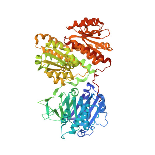
    Glutamine fructose-6-phosphate amidotransferase (GFAT) is the key enzyme in the hexosamine pathway (HP) that produces uridine 5'-diphospho-N-acetyl-D-glucosamine (UDP-GlcNAc), linking energy metabolism with posttranslational protein glycosylation. In Caenorhabditis elegans, we previously identified gfat-1 gain-of-function mutations that elevate UDP-GlcNAc levels, improve protein homeostasis, and extend lifespan. GFAT is highly conserved, but the gain-of-function mechanism and its relevance in mammalian cells remained unclear. Here, we present the full-length crystal structure of human GFAT-1 in complex with various ligands and with important mutations. UDP-GlcNAc directly interacts with GFAT-1, inhibiting catalytic activity. The longevity-associated G451E variant shows drastically reduced sensitivity to UDP-GlcNAc inhibition in enzyme activity assays. Our structural and functional data point to a critical role of the interdomain linker in UDP-GlcNAc inhibition. In mammalian cells, the G451E variant potently activates the HP. Therefore, GFAT-1 gain-of-function through loss of feedback inhibition constitutes a potential target for the treatment of age-related proteinopathies.


  • Organizational Affiliation
    • Max Planck Institute for Biology of Ageing, 50931, Cologne, Germany.

Macromolecule Content 

  • Total Structure Weight: 157.32 kDa 
  • Atom Count: 10,565 
  • Modeled Residue Count: 1,315 
  • Deposited Residue Count: 1,374 
  • Unique protein chains: 1

Macromolecules

Find similar proteins by:|  3D Structure
Entity ID: 1
MoleculeChains  Sequence LengthOrganismDetailsImage
Glutamine--fructose-6-phosphate aminotransferase [isomerizing] 1
A, B
687Homo sapiensMutation(s): 0 
Gene Names: GFPT1GFATGFPT
EC: 2.6.1.16
UniProt & NIH Common Fund Data Resources
Find proteins for Q06210 (Homo sapiens)
Explore Q06210 
Go to UniProtKB:  Q06210
PHAROS:  Q06210
GTEx:  ENSG00000198380 
Entity Groups
Sequence Clusters30% Identity50% Identity70% Identity90% Identity95% Identity100% Identity
UniProt GroupQ06210
Sequence Annotations
Expand
Reference Sequence

Small Molecules

Ligands 4 Unique
IDChains Name / Formula / InChI Key2D Diagram3D Interactions
UD2
(Subject of Investigation/LOI)

Query on UD2


Download:Ideal Coordinates CCD File
E [auth A],
H [auth B]
URIDINE-DIPHOSPHATE-N-ACETYLGALACTOSAMINE
C17 H27 N3 O17 P2
LFTYTUAZOPRMMI-NESSUJCYSA-N
G6Q

Query on G6Q


Download:Ideal Coordinates CCD File
C [auth A],
G [auth B]
GLUCOSE-6-PHOSPHATE
C6 H13 O9 P
VFRROHXSMXFLSN-SLPGGIOYSA-N
GLU

Query on GLU


Download:Ideal Coordinates CCD File
D [auth A]GLUTAMIC ACID
C5 H9 N O4
WHUUTDBJXJRKMK-VKHMYHEASA-N
MG

Query on MG


Download:Ideal Coordinates CCD File
F [auth A],
I [auth B]
MAGNESIUM ION
Mg
JLVVSXFLKOJNIY-UHFFFAOYSA-N

Experimental Data & Validation

Experimental Data

  • Method: X-RAY DIFFRACTION
  • Resolution: 2.48 Å
  • R-Value Free:  0.222 (Depositor), 0.229 (DCC) 
  • R-Value Work:  0.189 (Depositor), 0.194 (DCC) 
  • R-Value Observed: 0.190 (Depositor) 
Space Group: P 41 21 2
Unit Cell:
Length ( Å )Angle ( ˚ )
a = 151.995α = 90
b = 151.995β = 90
c = 165.803γ = 90
Software Package:
Software NamePurpose
PHENIXrefinement
PDB_EXTRACTdata extraction
XDSdata reduction
XDSdata scaling
PHENIXphasing

Structure Validation

View Full Validation Report


Ligand Structure Quality Assessment 


Entry History 

& Funding Information

Deposition Data


Funding OrganizationLocationGrant Number
German Federal Ministry for Education and ResearchGermany01GQ1423A
European CommissionGermanyERC-2014-StG-640254-MetAGEn
European UnionGermanyiNEXT, Horizon 2020

Revision History  (Full details and data files)

  • Version 1.0: 2020-01-15
    Type: Initial release
  • Version 1.1: 2020-02-19
    Changes: Database references
  • Version 1.2: 2024-01-24
    Changes: Data collection, Database references, Derived calculations, Refinement description