6SSD | pdb_00006ssd

Transaminase with PLP bound


Experimental Data Snapshot

  • Method: X-RAY DIFFRACTION
  • Resolution: 1.18 Å
  • R-Value Free: 
    0.129 (Depositor), 0.154 (DCC) 
  • R-Value Work: 
    0.111 (Depositor) 
  • R-Value Observed: 
    0.112 (Depositor) 

wwPDB Validation 3D Report Full Report

Validation slider image for 6SSD

Ligand Structure Quality Assessment 


This is version 1.1 of the entry. See complete history

Literature

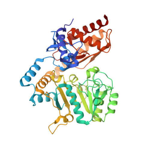
PMP-diketopiperazine adducts form at the active site of a PLP dependent enzyme involved in formycin biosynthesis.

Gao, S.Liu, H.de Crecy-Lagard, V.Zhu, W.Richards, N.G.J.Naismith, J.H.

(2019) Chem Commun (Camb) 55: 14502-14505

  • DOI: https://doi.org/10.1039/c9cc06975e
  • Primary Citation Related Structures: 
    6SSD, 6SSE, 6SSF, 6SSG

  • PubMed Abstract: 

    ForI is a PLP-dependent enzyme from the biosynthetic pathway of the C-nucleoside antibiotic formycin. Cycloserine is thought to inhibit PLP-dependent enzymes by irreversibly forming a PMP-isoxazole. We now report that ForI forms novel PMP-diketopiperazine derivatives following incubation with both d and l cycloserine. This unexpected result suggests chemical diversity in the chemistry of cycloserine inhibition.


  • Organizational Affiliation
    • Research Complex at Harwell, Didcot, OX11 0FA, UK.

Macromolecule Content 

  • Total Structure Weight: 44.9 kDa 
  • Atom Count: 4,050 
  • Modeled Residue Count: 424 
  • Deposited Residue Count: 424 
  • Unique protein chains: 1

Macromolecules

Find similar proteins by:|  3D Structure
Entity ID: 1
MoleculeChains  Sequence LengthOrganismDetailsImage
ForI-PLP424Streptomyces kaniharaensisMutation(s): 0 
UniProt
Find proteins for A0A6I8WFW1 (Streptomyces kaniharaensis)
Explore A0A6I8WFW1 
Go to UniProtKB:  A0A6I8WFW1
Entity Groups
Sequence Clusters30% Identity50% Identity70% Identity90% Identity95% Identity100% Identity
UniProt GroupA0A6I8WFW1
Sequence Annotations
Expand
Reference Sequence

Experimental Data & Validation

Experimental Data

  • Method: X-RAY DIFFRACTION
  • Resolution: 1.18 Å
  • R-Value Free:  0.129 (Depositor), 0.154 (DCC) 
  • R-Value Work:  0.111 (Depositor) 
  • R-Value Observed: 0.112 (Depositor) 
Space Group: P 43 21 2
Unit Cell:
Length ( Å )Angle ( ˚ )
a = 92.3α = 90
b = 92.3β = 90
c = 118.8γ = 90
Software Package:
Software NamePurpose
REFMACrefinement
xia2data scaling
PDB_EXTRACTdata extraction
xia2data reduction
PHASERphasing

Structure Validation

View Full Validation Report


Ligand Structure Quality Assessment 


Entry History 

Deposition Data

Revision History  (Full details and data files)

  • Version 1.0: 2020-01-15
    Type: Initial release
  • Version 1.1: 2025-04-09
    Changes: Data collection, Database references, Structure summary