Structural and functional characterization of C0021158, a high-affinity monoclonal antibody that inhibits Arginase 2 function via a novel non-competitive mechanism of action.
Austin, M., Burschowsky, D., Chan, D.T.Y., Jenkinson, L., Haynes, S., Diamandakis, A., Seewooruthun, C., Addyman, A., Fiedler, S., Ryman, S., Whitehouse, J., Slater, L.H., Hadjinicolaou, A.V., Gileadi, U., Gowans, E., Shibata, Y., Barnard, M., Kaserer, T., Sharma, P., Luheshi, N.M., Wilkinson, R.W., Vaughan, T.J., Holt, S.V., Cerundolo, V., Carr, M.D., Groves, M.A.T.(null) MAbs 12: 1801230-1801230
- PubMed: 32880207 Search on PubMedSearch on PubMed Central
- DOI: https://doi.org/10.1080/19420862.2020.1801230
- Primary Citation Related Structures:
6SRV, 6SRX, 6SS0, 6SS2, 6SS4, 6TUL - PubMed Abstract:
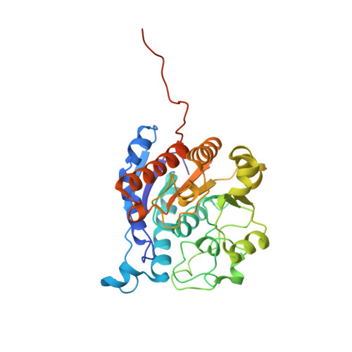
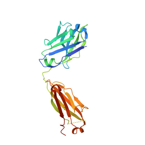
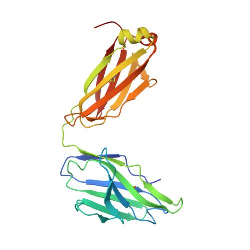
Arginase 2 (ARG2) is a binuclear manganese metalloenzyme that catalyzes the hydrolysis of L-arginine. The dysregulated expression of ARG2 within specific tumor microenvironments generates an immunosuppressive niche that effectively renders the tumor 'invisible' to the host's immune system. Increased ARG2 expression leads to a concomitant depletion of local L-arginine levels, which in turn leads to suppression of anti-tumor T-cell-mediated immune responses. Here we describe the isolation and characterization of a high affinity antibody (C0021158) that inhibits ARG2 enzymatic function completely, effectively restoring T-cell proliferation in vitro . Enzyme kinetic studies confirmed that C0021158 exhibits a noncompetitive mechanism of action, inhibiting ARG2 independently of L-arginine concentrations. To elucidate C0021158's inhibitory mechanism at a structural level, the co-crystal structure of the Fab in complex with trimeric ARG2 was solved. C0021158's epitope was consequently mapped to an area some distance from the enzyme's substrate binding cleft, indicating an allosteric mechanism was being employed. Following C0021158 binding, distinct regions of ARG2 undergo major conformational changes. Notably, the backbone structure of a surface-exposed loop is completely rearranged, leading to the formation of a new short helix structure at the Fab-ARG2 interface. Moreover, this large-scale structural remodeling at ARG2's epitope translates into more subtle changes within the enzyme's active site. An arginine residue at position 39 is reoriented inwards, sterically impeding the binding of L-arginine. Arg39 is also predicted to alter the p K A of a key catalytic histidine residue at position 160, further attenuating ARG2's enzymatic function. In silico molecular docking simulations predict that L-arginine is unable to bind effectively when antibody is bound, a prediction supported by isothermal calorimetry experiments using an L-arginine mimetic. Specifically, targeting ARG2 in the tumor microenvironment through the application of C0021158, potentially in combination with standard chemotherapy regimens or alternate immunotherapies, represents a potential new strategy to target immune cold tumors.
- Cancer Research UK AstraZeneca Antibody Alliance Laboratory , Cambridge, UK.
Organizational Affiliation: