6SCX

Crystal structure of the catalytic domain of human NUDT12 in complex with 7-methyl-guanosine-5'-triphosphate


Experimental Data Snapshot

  • Method: X-RAY DIFFRACTION
  • Resolution: 2.92 Å
  • R-Value Free: 0.260 
  • R-Value Work: 0.232 
  • R-Value Observed: 0.233 

wwPDB Validation   3D Report Full Report


Ligand Structure Quality Assessment 


This is version 1.1 of the entry. See complete history


Literature

Decapping Enzyme NUDT12 Partners with BLMH for Cytoplasmic Surveillance of NAD-Capped RNAs.

Wu, H.Li, L.Chen, K.M.Homolka, D.Gos, P.Fleury-Olela, F.McCarthy, A.A.Pillai, R.S.

(2019) Cell Rep 29: 4422-4434.e13

  • DOI: https://doi.org/10.1016/j.celrep.2019.11.108
  • Primary Citation of Related Structures:  
    6SCX

  • PubMed Abstract: 

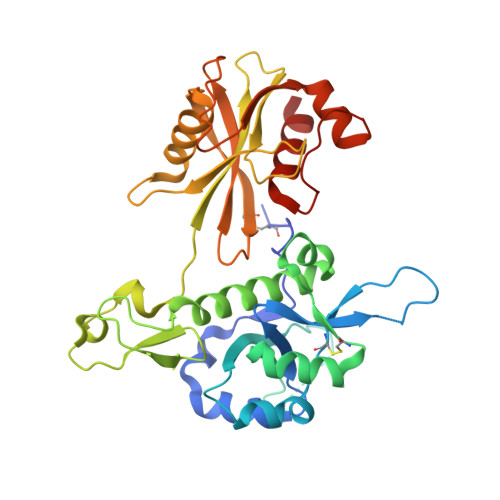
    RNA polymerase II transcripts receive a protective 5',5'-triphosphate-linked 7-methylguanosine (m 7 G) cap, and its removal by decapping enzymes like DCP2 is critical for initiation of RNA decay. Alternative RNA caps can be acquired when transcription initiation uses metabolites like nicotinamide adenine dinucleotide (NAD), generating NAD-RNAs. Here, we identify human NUDT12 as a cytosolic NAD-RNA decapping enzyme. NUDT12 is active only as homodimers, with each monomer contributing to creation of the two functional catalytic pockets. We identify an ∼600-kDa dodecamer complex between bleomycin hydrolase (BLMH) and NUDT12, with BLMH being required for localization of NUDT12 to a few discrete cytoplasmic granules that are distinct from P-bodies. Both proteins downregulate gene expression when artificially tethered to a reporter RNA in vivo. Furthermore, loss of Nudt12 results in a significant upregulation of circadian clock transcripts in mouse liver. Overall, our study points to a physiological role for NUDT12 in the cytosolic surveillance of NAD-RNAs.


  • Organizational Affiliation

    Department of Molecular Biology, Science III, University of Geneva, 30 Quai Ernest-Ansermet, 1211 Geneva 4, Switzerland.


Macromolecules
Find similar proteins by:  (by identity cutoff)  |  3D Structure
Entity ID: 1
MoleculeChains Sequence LengthOrganismDetailsImage
Peroxisomal NADH pyrophosphatase NUDT12
A, B, C
352Homo sapiensMutation(s): 0 
Gene Names: NUDT12
EC: 3.6.1.22
UniProt & NIH Common Fund Data Resources
Find proteins for Q9BQG2 (Homo sapiens)
Explore Q9BQG2 
Go to UniProtKB:  Q9BQG2
PHAROS:  Q9BQG2
GTEx:  ENSG00000112874 
Entity Groups  
Sequence Clusters30% Identity50% Identity70% Identity90% Identity95% Identity100% Identity
UniProt GroupQ9BQG2
Sequence Annotations
Expand
  • Reference Sequence
Small Molecules
Ligands 2 Unique
IDChains Name / Formula / InChI Key2D Diagram3D Interactions
MGP
Query on MGP

Download Ideal Coordinates CCD File 
H [auth A],
L [auth B],
O [auth C]
7-METHYL-GUANOSINE-5'-TRIPHOSPHATE
C11 H19 N5 O14 P3
DKVRNHPCAOHRSI-KQYNXXCUSA-O
CD
Query on CD

Download Ideal Coordinates CCD File 
D [auth A]
E [auth A]
F [auth A]
G [auth A]
I [auth B]
D [auth A],
E [auth A],
F [auth A],
G [auth A],
I [auth B],
J [auth B],
K [auth B],
M [auth C],
N [auth C]
CADMIUM ION
Cd
WLZRMCYVCSSEQC-UHFFFAOYSA-N
Experimental Data & Validation

Experimental Data

Unit Cell:
Length ( Å )Angle ( ˚ )
a = 229.859α = 90
b = 229.859β = 90
c = 159.012γ = 120
Software Package:
Software NamePurpose
BUSTERrefinement
PDB_EXTRACTdata extraction
Cootmodel building
PHASERphasing
pointlessdata scaling
XDSdata reduction
autoPROCdata scaling
STARANISOdata scaling
MxCuBEdata collection

Structure Validation

View Full Validation Report


Ligand Structure Quality Assessment 


Entry History 

Deposition Data

Revision History  (Full details and data files)

  • Version 1.0: 2020-01-08
    Type: Initial release
  • Version 1.1: 2024-01-24
    Changes: Data collection, Database references, Derived calculations, Refinement description