6SC9

dAb3/HOIP-RBR-HOIPIN-8


Experimental Data Snapshot

  • Method: X-RAY DIFFRACTION
  • Resolution: 2.47 Å
  • R-Value Free: 0.252 
  • R-Value Work: 0.210 
  • R-Value Observed: 0.212 

wwPDB Validation   3D Report Full Report


Ligand Structure Quality Assessment 


This is version 1.2 of the entry. See complete history


Literature

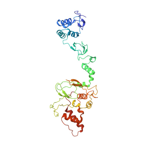
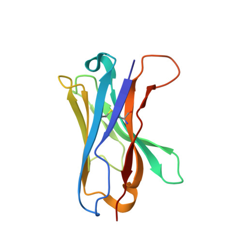
Single-Domain Antibodies as Crystallization Chaperones to Enable Structure-Based Inhibitor Development for RBR E3 Ubiquitin Ligases.

Tsai, Y.I.Johansson, H.Dixon, D.Martin, S.Chung, C.W.Clarkson, J.House, D.Rittinger, K.

(2020) Cell Chem Biol 27: 83

  • DOI: https://doi.org/10.1016/j.chembiol.2019.11.007
  • Primary Citation of Related Structures:  
    6SC5, 6SC6, 6SC7, 6SC8, 6SC9, 6T2J

  • PubMed Abstract: 

    Protein ubiquitination plays a key role in the regulation of cellular processes, and misregulation of the ubiquitin system is linked to many diseases. So far, development of tool compounds that target enzymes of the ubiquitin system has been slow and only a few specific inhibitors are available. Here, we report the selection of single-domain antibodies (single-dAbs) based on a human scaffold that recognize the catalytic domain of HOIP, a subunit of the multi-component E3 LUBAC and member of the RBR family of E3 ligases. Some of these dAbs affect ligase activity and provide mechanistic insight into the ubiquitin transfer mechanism of different E2-conjugating enzymes. Furthermore, we show that the co-crystal structure of a HOIP RBR/dAb complex serves as a robust platform for soaking of ligands that target the active site cysteine of HOIP, thereby providing easy access to structure-based ligand design for this important class of E3 ligases.


  • Organizational Affiliation

    Molecular Structure of Cell Signalling Laboratory, The Francis Crick Institute, 1 Midland Road, London NW1 1AT, UK.


Macromolecules
Find similar proteins by:  (by identity cutoff)  |  3D Structure
Entity ID: 1
MoleculeChains Sequence LengthOrganismDetailsImage
E3 ubiquitin-protein ligase RNF31376Homo sapiensMutation(s): 0 
Gene Names: RNF31ZIBRA
EC: 2.3.2.31
UniProt & NIH Common Fund Data Resources
Find proteins for Q96EP0 (Homo sapiens)
Explore Q96EP0 
Go to UniProtKB:  Q96EP0
PHAROS:  Q96EP0
GTEx:  ENSG00000092098 
Entity Groups  
Sequence Clusters30% Identity50% Identity70% Identity90% Identity95% Identity100% Identity
UniProt GroupQ96EP0
Sequence Annotations
Expand
  • Reference Sequence
Find similar proteins by:  (by identity cutoff)  |  3D Structure
Entity ID: 2
MoleculeChains Sequence LengthOrganismDetailsImage
Single domain antibody
B, C
120synthetic constructMutation(s): 0 
Entity Groups  
Sequence Clusters30% Identity50% Identity70% Identity90% Identity95% Identity100% Identity
Sequence Annotations
Expand
  • Reference Sequence
Small Molecules
Ligands 4 Unique
IDChains Name / Formula / InChI Key2D Diagram3D Interactions
L68 (Subject of Investigation/LOI)
Query on L68

Download Ideal Coordinates CCD File 
D [auth A]2-[3-[2,6-bis(fluoranyl)-4-(1~{H}-pyrazol-4-yl)phenyl]-3-oxidanylidene-prop-1-enyl]-4-(1-methylpyrazol-4-yl)benzoic acid
C23 H16 F2 N4 O3
UMAKCENGRSEXNB-HWKANZROSA-N
SO4
Query on SO4

Download Ideal Coordinates CCD File 
M [auth A],
O [auth B]
SULFATE ION
O4 S
QAOWNCQODCNURD-UHFFFAOYSA-L
ZN
Query on ZN

Download Ideal Coordinates CCD File 
E [auth A]
F [auth A]
G [auth A]
H [auth A]
I [auth A]
E [auth A],
F [auth A],
G [auth A],
H [auth A],
I [auth A],
J [auth A],
K [auth A],
L [auth A]
ZINC ION
Zn
PTFCDOFLOPIGGS-UHFFFAOYSA-N
CL
Query on CL

Download Ideal Coordinates CCD File 
N [auth A],
P [auth B]
CHLORIDE ION
Cl
VEXZGXHMUGYJMC-UHFFFAOYSA-M
Experimental Data & Validation

Experimental Data

  • Method: X-RAY DIFFRACTION
  • Resolution: 2.47 Å
  • R-Value Free: 0.252 
  • R-Value Work: 0.210 
  • R-Value Observed: 0.212 
  • Space Group: I 2 2 2
Unit Cell:
Length ( Å )Angle ( ˚ )
a = 66.127α = 90
b = 87.052β = 90
c = 241.538γ = 90
Software Package:
Software NamePurpose
REFMACrefinement
PHENIXrefinement
DIALSdata reduction
Aimlessdata scaling
PHENIXphasing

Structure Validation

View Full Validation Report


Ligand Structure Quality Assessment 


Entry History & Funding Information

Deposition Data


Funding OrganizationLocationGrant Number
The Francis Crick InstituteUnited Kingdom--

Revision History  (Full details and data files)

  • Version 1.0: 2019-11-27
    Type: Initial release
  • Version 1.1: 2019-12-18
    Changes: Database references
  • Version 1.2: 2020-01-29
    Changes: Database references