6S8I | pdb_00006s8i

Structure of ZEBOV GP in complex with 3T0265 antibody


Experimental Data Snapshot

  • Method: ELECTRON MICROSCOPY
  • Resolution: 2.99 Å
  • Aggregation State: PARTICLE 
  • Reconstruction Method: SINGLE PARTICLE 

Starting Model: experimental
View more details

wwPDB Validation 3D Report Full Report

Validation slider image for 6S8I

This is version 2.1 of the entry. See complete history

Literature

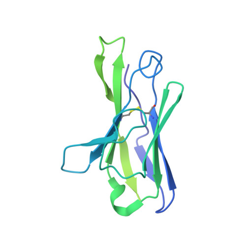
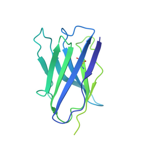
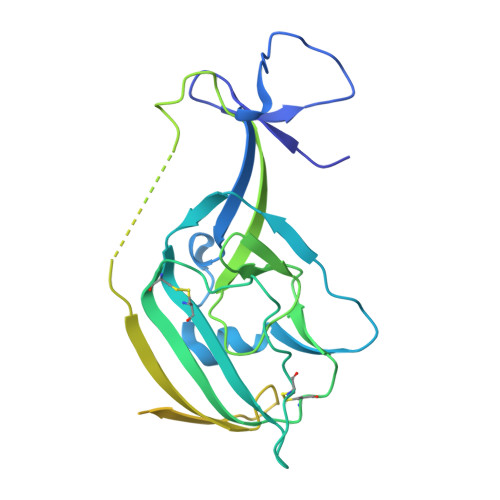
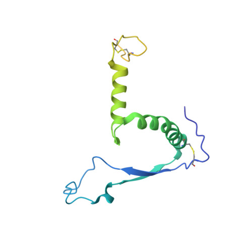
Structural Basis for a Convergent Immune Response against Ebola Virus.

Cohen-Dvashi, H.Zehner, M.Ehrhardt, S.Katz, M.Elad, N.Klein, F.Diskin, R.

(2020) Cell Host Microbe 27: 418-427.e4

  • DOI: https://doi.org/10.1016/j.chom.2020.01.007
  • Primary Citation Related Structures: 
    6S8D, 6S8I, 6S8J

  • PubMed Abstract: 

    Ebola virus disease is a severe health problem in Africa. Vaccines that display the Zaire ebolavirus glycoprotein spike complex are a prime component for the effort to combat it. The V H 3-15/V λ 1-40-based class of antibodies was recently discovered to be a common response in individuals who received the Ebola virus vaccines. These antibodies display attractive properties, and thus likely contribute to the efficacy of the vaccines. Here, we use cryo-EM to elucidate how three V H 3-15/V λ 1-40 antibodies from different individuals target the virus and found a convergent mechanism against a partially conserved site on the spike complex. Our study rationalizes the selection of the V H 3-15/V λ 1-40 germline genes for specifically targeting this site and highlights Ebolavirus species-specific sequence divergences that may restrict breadth of V H 3-15/V λ 1-40-based humoral response. The results from this study could help develop improved immunization schemes and further enable the design of immunogens that would be efficacious against a broader set of Ebolavirus species.


  • Organizational Affiliation
    • Department of Structural Biology, Weizmann Institute of Science, Rehovot 76100, Israel.

Macromolecule Content 

  • Total Structure Weight: 305.96 kDa 
  • Atom Count: 12,066 
  • Modeled Residue Count: 1,566 
  • Deposited Residue Count: 2,799 
  • Unique protein chains: 4

Macromolecules

Find similar proteins by:|  3D Structure
Entity ID: 1
MoleculeChains  Sequence LengthOrganismDetailsImage
Light ChainA [auth L],
I [auth O],
J [auth U]
218Homo sapiensMutation(s): 0 
Find similar proteins by:|  3D Structure
Entity ID: 2
MoleculeChains  Sequence LengthOrganismDetailsImage
Heavy ChainB [auth H],
K [auth P],
L [auth Y]
224Homo sapiensMutation(s): 0 
Find similar proteins by:|  3D Structure
Entity ID: 3
MoleculeChains  Sequence LengthOrganismDetailsImage
Enveloped Glycoprotein 1C [auth A],
G [auth C],
H [auth E]
323Ebola virusMutation(s): 0 
UniProt
Find proteins for Q05320 (Zaire ebolavirus (strain Mayinga-76))
Explore Q05320 
Go to UniProtKB:  Q05320
Entity Groups
Sequence Clusters30% Identity50% Identity70% Identity90% Identity95% Identity100% Identity
UniProt GroupQ05320
Sequence Annotations
Expand
Reference Sequence
Find similar proteins by:|  3D Structure
Entity ID: 4
MoleculeChains  Sequence LengthOrganismDetailsImage
Envelope glycoproteinD [auth B],
E [auth D],
F
168Ebola virusMutation(s): 0 
Gene Names: 
UniProt
Find proteins for Q05320 (Zaire ebolavirus (strain Mayinga-76))
Explore Q05320 
Go to UniProtKB:  Q05320
Entity Groups
Sequence Clusters30% Identity50% Identity70% Identity90% Identity95% Identity100% Identity
UniProt GroupQ05320
Glycosylation
Glycosylation Sites: 1
Sequence Annotations
Expand
Reference Sequence

Oligosaccharides

Help  
Entity ID: 5
MoleculeChains Length2D Diagram GlycosylationD Interactions
2-acetamido-2-deoxy-beta-D-glucopyranose-(1-4)-2-acetamido-2-deoxy-beta-D-glucopyranoseM [auth G],
N [auth I],
O [auth J]
2N-Glycosylation
Glycosylation Resources
GlyTouCan: G42666HT
GlyCosmos: G42666HT
GlyGen: G42666HT

Experimental Data & Validation

Experimental Data

  • Method: ELECTRON MICROSCOPY
  • Resolution: 2.99 Å
  • Aggregation State: PARTICLE 
  • Reconstruction Method: SINGLE PARTICLE 
EM Software:
TaskSoftware PackageVersion
MODEL REFINEMENTPHENIX3297
RECONSTRUCTIONcryoSPARCV2

Structure Validation

View Full Validation Report


Entry History 

Deposition Data

Revision History  (Full details and data files)

  • Version 1.0: 2020-02-12
    Type: Initial release
  • Version 1.1: 2020-02-26
    Changes: Database references
  • Version 1.2: 2020-03-25
    Changes: Database references
  • Version 2.0: 2020-07-29
    Type: Remediation
    Reason: Carbohydrate remediation
    Changes: Atomic model, Data collection, Derived calculations, Structure summary
  • Version 2.1: 2024-10-23
    Changes: Data collection, Database references, Refinement description, Structure summary