Structural and biochemical evaluation of bisubstrate inhibitors of protein arginine N-methyltransferases PRMT1 and CARM1 (PRMT4).
Gunnell, E.A., Al-Noori, A., Muhsen, U., Davies, C.C., Dowden, J., Dreveny, I.(2020) Biochem J 477: 787-800
- PubMed: 32011657 Search on PubMedSearch on PubMed Central
- DOI: https://doi.org/10.1042/BCJ20190826
- Primary Citation Related Structures:
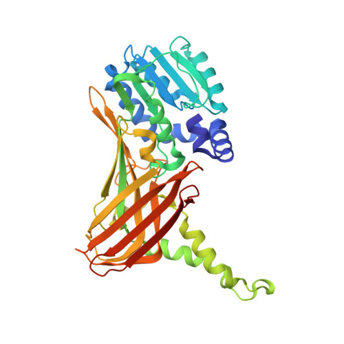
6S70, 6S71, 6S74, 6S77, 6S79, 6S7A, 6S7B, 6S7C - PubMed Abstract:
Attenuating the function of protein arginine methyltransferases (PRMTs) is an objective for the investigation and treatment of several diseases including cardiovascular disease and cancer. Bisubstrate inhibitors that simultaneously target binding sites for arginine substrate and the cofactor (S-adenosylmethionine (SAM)) have potential utility, but structural information on their binding is required for their development. Evaluation of bisubstrate inhibitors featuring an isosteric guanidine replacement with two prominent enzymes PRMT1 and CARM1 (PRMT4) by isothermal titration calorimetry (ITC), activity assays and crystallography are reported. Key findings are that 2-aminopyridine is a viable replacement for guanidine, providing an inhibitor that binds more strongly to CARM1 than PRMT1. Moreover, a residue around the active site that differs between CARM1 (Asn-265) and PRMT1 (Tyr-160) is identified that affects the side chain conformation of the catalytically important neighbouring glutamate in the crystal structures. Mutagenesis data supports its contribution to the difference in binding observed for this inhibitor. Structures of CARM1 in complex with a range of seven inhibitors reveal the binding modes and show that inhibitors with an amino acid terminus adopt a single conformation whereas the electron density for equivalent amine-bearing inhibitors is consistent with preferential binding in two conformations. These findings inform the molecular basis of CARM1 ligand binding and identify differences between CARM1 and PRMT1 that can inform drug discovery efforts.
- School of Chemistry, University of Nottingham, Nottingham NG7 2RD, U.K.
Organizational Affiliation: