


First crystal structure of parasitic PEX14 in complex with a fragment molecule 1H-indole-7-carboxylic acid
Hassaan, E., Heine, A., Klebe, G.To be published.
Experimental Data Snapshot
Entity ID: 1 | |||||
|---|---|---|---|---|---|
| Molecule | Chains | Sequence Length | Organism | Details | Image |
| Peroxin 14 | 69 | Trypanosoma brucei brucei | Mutation(s): 1 Gene Names: PEX14 | 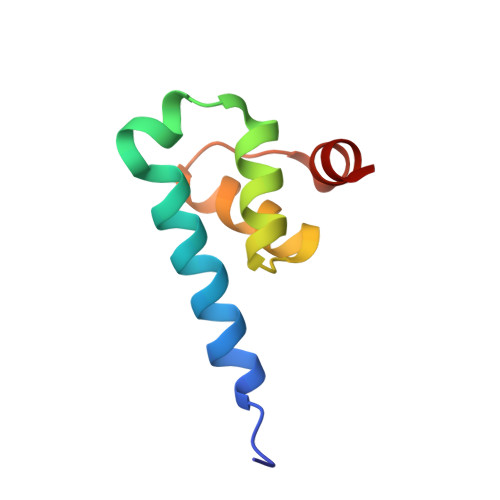 | |
UniProt | |||||
Entity Groups | |||||
| Sequence Clusters | 30% Identity50% Identity70% Identity90% Identity95% Identity100% Identity | ||||
| UniProt Group | Q38CL4 | ||||
Sequence AnnotationsExpand | |||||
Reference Sequence | |||||
| Ligands 4 Unique | |||||
|---|---|---|---|---|---|
| ID | Chains | Name / Formula / InChI Key | 2D Diagram | 3D Interactions | |
| KXQ (Subject of Investigation/LOI) Download:Ideal Coordinates CCD File | E [auth A] | 1~{H}-indole-7-carboxylic acid C9 H7 N O2 IPDOBVFESNNYEE-UHFFFAOYSA-N |  | ||
| SO4 Download:Ideal Coordinates CCD File | B [auth A], C [auth A], D [auth A] | SULFATE ION O4 S QAOWNCQODCNURD-UHFFFAOYSA-L |  | ||
| GOL Download:Ideal Coordinates CCD File | F [auth A] | GLYCEROL C3 H8 O3 PEDCQBHIVMGVHV-UHFFFAOYSA-N |  | ||
| DMS Download:Ideal Coordinates CCD File | G [auth A] | DIMETHYL SULFOXIDE C2 H6 O S IAZDPXIOMUYVGZ-UHFFFAOYSA-N |  | ||
| Length ( Å ) | Angle ( ˚ ) |
|---|---|
| a = 43.306 | α = 90 |
| b = 43.306 | β = 90 |
| c = 80.942 | γ = 90 |
| Software Name | Purpose |
|---|---|
| PHENIX | refinement |
| PHASER | phasing |
| Coot | model building |
| XDS | data scaling |
| XDS | data reduction |
| Funding Organization | Location | Grant Number |
|---|---|---|
| European Commission | Germany | -- |


