Pharmacological targeting of MTHFD2 suppresses acute myeloid leukemia by inducing thymidine depletion and replication stress.
Bonagas, N., Gustafsson, N.M.S., Henriksson, M., Marttila, P., Gustafsson, R., Wiita, E., Borhade, S., Green, A.C., Vallin, K.S.A., Sarno, A., Svensson, R., Gokturk, C., Pham, T., Jemth, A.S., Loseva, O., Cookson, V., Kiweler, N., Sandberg, L., Rasti, A., Unterlass, J.E., Haraldsson, M., Andersson, Y., Scaletti, E.R., Bengtsson, C., Paulin, C.B.J., Sanjiv, K., Abdurakhmanov, E., Pudelko, L., Kunz, B., Desroses, M., Iliev, P., Farnegardh, K., Kramer, A., Garg, N., Michel, M., Haggblad, S., Jarvius, M., Kalderen, C., Jensen, A.B., Almlof, I., Karsten, S., Zhang, S.M., Haggblad, M., Eriksson, A., Liu, J., Glinghammar, B., Nekhotiaeva, N., Klingegard, F., Koolmeister, T., Martens, U., Llona-Minguez, S., Moulson, R., Nordstrom, H., Parrow, V., Dahllund, L., Sjoberg, B., Vargas, I.L., Vo, D.D., Wannberg, J., Knapp, S., Krokan, H.E., Arvidsson, P.I., Scobie, M., Meiser, J., Stenmark, P., Berglund, U.W., Homan, E.J., Helleday, T.(2022) Nat Cancer 3: 156-172
- PubMed: 35228749 Search on PubMedSearch on PubMed Central
- DOI: https://doi.org/10.1038/s43018-022-00331-y
- Primary Citation Related Structures:
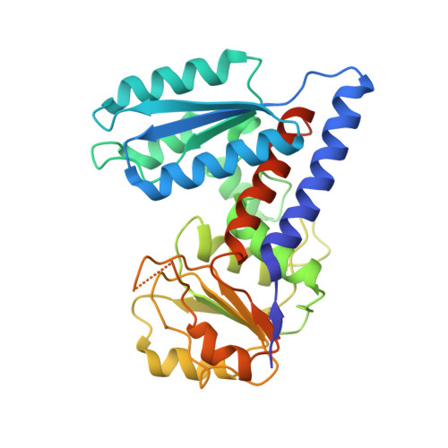
6S4A, 6S4E, 6S4F - PubMed Abstract:
The folate metabolism enzyme MTHFD2 (methylenetetrahydrofolate dehydrogenase/cyclohydrolase) is consistently overexpressed in cancer but its roles are not fully characterized, and current candidate inhibitors have limited potency for clinical development. In the present study, we demonstrate a role for MTHFD2 in DNA replication and genomic stability in cancer cells, and perform a drug screen to identify potent and selective nanomolar MTHFD2 inhibitors; protein cocrystal structures demonstrated binding to the active site of MTHFD2 and target engagement. MTHFD2 inhibitors reduced replication fork speed and induced replication stress followed by S-phase arrest and apoptosis of acute myeloid leukemia cells in vitro and in vivo, with a therapeutic window spanning four orders of magnitude compared with nontumorigenic cells. Mechanistically, MTHFD2 inhibitors prevented thymidine production leading to misincorporation of uracil into DNA and replication stress. Overall, these results demonstrate a functional link between MTHFD2-dependent cancer metabolism and replication stress that can be exploited therapeutically with this new class of inhibitors.
- Science for Life Laboratory, Department of Oncology-Pathology, Karolinska Institutet, Solna, Sweden.
Organizational Affiliation: