6RUU

Pseudokinase domain of human IRAK3


Experimental Data Snapshot

  • Method: X-RAY DIFFRACTION
  • Resolution: 2.95 Å
  • R-Value Free: 0.260 
  • R-Value Work: 0.228 
  • R-Value Observed: 0.230 

wwPDB Validation   3D Report Full Report


This is version 1.4 of the entry. See complete history


Literature

Dimeric Structure of the Pseudokinase IRAK3 Suggests an Allosteric Mechanism for Negative Regulation.

Lange, S.M.Nelen, M.I.Cohen, P.Kulathu, Y.

(2021) Structure 29: 238

  • DOI: https://doi.org/10.1016/j.str.2020.11.004
  • Primary Citation of Related Structures:  
    6RUU

  • PubMed Abstract: 

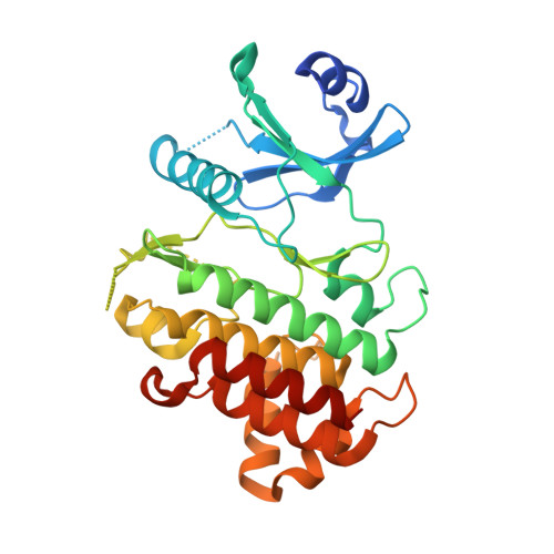
    Interleukin-1 receptor associated kinases (IRAKs) are key players in innate immune signaling that mediate the host response to pathogens. In contrast to the active kinases IRAK1 and IRAK4, IRAK2 and IRAK3 are pseudokinases lacking catalytic activity and their functions are poorly understood. IRAK3 is thought to be a negative regulator of innate immune signaling and mutations in IRAK3 are associated with asthma and cancer. Here, we report the crystal structure of the human IRAK3 pseudokinase domain in a closed, pseudoactive conformation. IRAK3 dimerizes in a unique way through a head-to-head arrangement not observed in any other kinases. Multiple conserved cysteine residues imply a potential redox control of IRAK3 conformation and dimerization. By analyzing asthma-associated mutations, we identify an evolutionarily conserved surface on IRAK3 that could form an interaction interface with IRAK4, suggesting a model for the negative regulation of IRAK4 by IRAK3.


  • Organizational Affiliation

    MRC Protein Phosphorylation and Ubiquitylation Unit, Sir James Black Centre, Dow Street, Dundee, Scotland DD1 5EH, UK.


Macromolecules
Find similar proteins by:  (by identity cutoff)  |  3D Structure
Entity ID: 1
MoleculeChains Sequence LengthOrganismDetailsImage
Interleukin-1 receptor-associated kinase 3
A, B, C
315Homo sapiensMutation(s): 1 
Gene Names: IRAK3
EC: 2.7.11.1
UniProt & NIH Common Fund Data Resources
Find proteins for Q9Y616 (Homo sapiens)
Explore Q9Y616 
Go to UniProtKB:  Q9Y616
PHAROS:  Q9Y616
GTEx:  ENSG00000090376 
Entity Groups  
Sequence Clusters30% Identity50% Identity70% Identity90% Identity95% Identity100% Identity
UniProt GroupQ9Y616
Sequence Annotations
Expand
  • Reference Sequence
Small Molecules
Ligands 3 Unique
IDChains Name / Formula / InChI Key2D Diagram3D Interactions
HG
Query on HG

Download Ideal Coordinates CCD File 
D [auth A]
E [auth A]
L [auth B]
O [auth C]
P [auth C]
D [auth A],
E [auth A],
L [auth B],
O [auth C],
P [auth C],
Q [auth C],
R [auth C],
S [auth C]
MERCURY (II) ION
Hg
BQPIGGFYSBELGY-UHFFFAOYSA-N
SO4
Query on SO4

Download Ideal Coordinates CCD File 
H [auth A]
I [auth A]
J [auth A]
K [auth A]
M [auth B]
H [auth A],
I [auth A],
J [auth A],
K [auth A],
M [auth B],
N [auth B],
U [auth C],
V [auth C]
SULFATE ION
O4 S
QAOWNCQODCNURD-UHFFFAOYSA-L
GOL
Query on GOL

Download Ideal Coordinates CCD File 
F [auth A],
G [auth A],
T [auth C]
GLYCEROL
C3 H8 O3
PEDCQBHIVMGVHV-UHFFFAOYSA-N
Experimental Data & Validation

Experimental Data

  • Method: X-RAY DIFFRACTION
  • Resolution: 2.95 Å
  • R-Value Free: 0.260 
  • R-Value Work: 0.228 
  • R-Value Observed: 0.230 
  • Space Group: P 21 21 2
Unit Cell:
Length ( Å )Angle ( ˚ )
a = 53.55α = 90
b = 167.16β = 90
c = 179.51γ = 90
Software Package:
Software NamePurpose
PHENIXrefinement
MxCuBEdata collection
XDSdata reduction
Aimlessdata scaling
STARANISOdata scaling
MoRDaphasing
BALBESphasing
PARROTphasing
Cootmodel building

Structure Validation

View Full Validation Report


Entry History 

Deposition Data

Revision History  (Full details and data files)

  • Version 1.0: 2020-09-09
    Type: Initial release
  • Version 1.1: 2020-12-02
    Changes: Database references
  • Version 1.2: 2020-12-09
    Changes: Database references
  • Version 1.3: 2021-03-17
    Changes: Database references
  • Version 1.4: 2024-01-24
    Changes: Data collection, Database references, Refinement description