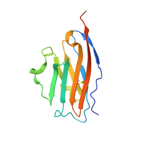
6RPG

Structure of human Programmed cell death 1 ligand 1 (PD-L1) with inhibitor


Experimental Data Snapshot

  • Method: X-RAY DIFFRACTION
  • Resolution: 2.70 Å
  • R-Value Free: 0.281 
  • R-Value Work: 0.248 
  • R-Value Observed: 0.251 

Starting Model: experimental
View more details

wwPDB Validation   3D Report Full Report


Ligand Structure Quality Assessment 


This is version 1.3 of the entry. See complete history


Literature

Design, Synthesis, Evaluation, and Structural Studies ofC2-Symmetric Small Molecule Inhibitors of Programmed Cell Death-1/Programmed Death-Ligand 1 Protein-Protein Interaction.

Basu, S.Yang, J.Xu, B.Magiera-Mularz, K.Skalniak, L.Musielak, B.Kholodovych, V.Holak, T.A.Hu, L.

(2019) J Med Chem 62: 7250-7263

  • DOI: https://doi.org/10.1021/acs.jmedchem.9b00795
  • Primary Citation of Related Structures:  
    6RPG

  • PubMed Abstract: 

    A series of C 2 -symmetric inhibitors was designed and evaluated for inhibitory activity against the programmed cell death-1/programmed death-ligand 1(PD-1/PD-L1) protein-protein interaction (PPI) in a homogenous time-resolved fluorescence (HTRF) assay and PD-1 signaling in cell-based coculture assays. C 2 -symmetric inhibitors 2a (LH1306) and 2b (LH1307) exhibited IC 50 values of 25 and 3.0 nM, respectively, in the HTRF assay. While 2a was ∼3.8-fold more potent than previously reported inhibitor 1a , 2b could not be differentiated from 1b due to their high potency and the limit of our HTRF assay conditions. In one cell-based coculture PD-1 signaling assay, 2a and 2b were 8.2- and 2.8-fold more potent in inhibiting PD-1 signaling than 1a and 1b , respectively. NMR and X-ray cocrystal structural studies provided more structural insights into the interaction between 2b and PD-L1; 2b binds to PD-L1 at the PD-1 binding site and induces the formation of a more symmetrically arranged PD-L1 homodimer than that previously reported for other inhibitors.


  • Organizational Affiliation

    Department of Organic Chemistry, Faculty of Chemistry , Jagiellonian University , Gronostajowa 2 , 30-387 Krakow , Poland.


Macromolecules
Find similar proteins by:  (by identity cutoff)  |  3D Structure
Entity ID: 1
MoleculeChains Sequence LengthOrganismDetailsImage
Programmed cell death 1 ligand 1
A, B
128Homo sapiensMutation(s): 0 
Gene Names: CD274B7H1PDCD1L1PDCD1LG1PDL1
UniProt & NIH Common Fund Data Resources
Find proteins for Q9NZQ7 (Homo sapiens)
Explore Q9NZQ7 
Go to UniProtKB:  Q9NZQ7
PHAROS:  Q9NZQ7
GTEx:  ENSG00000120217 
Entity Groups  
Sequence Clusters30% Identity50% Identity70% Identity90% Identity95% Identity100% Identity
UniProt GroupQ9NZQ7
Sequence Annotations
Expand
  • Reference Sequence
Small Molecules
Ligands 1 Unique
IDChains Name / Formula / InChI Key2D Diagram3D Interactions
KDW
Query on KDW

Download Ideal Coordinates CCD File 
C [auth B]~{N}-[2-[[4-[[3-[3-[[4-[(2-acetamidoethylamino)methyl]-5-[(5-cyanopyridin-3-yl)methoxy]-2-methyl-phenoxy]methyl]-2-methyl-phenyl]-2-methyl-phenyl]methoxy]-2-[(5-cyanopyridin-3-yl)methoxy]-5-methyl-phenyl]methylamino]ethyl]ethanamide
C54 H58 N8 O6
RNCINFJGGSQEHN-UHFFFAOYSA-N
Binding Affinity Annotations 
IDSourceBinding Affinity
KDW BindingDB:  6RPG IC50: 3 (nM) from 1 assay(s)
EC50: min: 79, max: 763 (nM) from 2 assay(s)
Experimental Data & Validation

Experimental Data

  • Method: X-RAY DIFFRACTION
  • Resolution: 2.70 Å
  • R-Value Free: 0.281 
  • R-Value Work: 0.248 
  • R-Value Observed: 0.251 
  • Space Group: P 31 2 1
Unit Cell:
Length ( Å )Angle ( ˚ )
a = 75.894α = 90
b = 75.894β = 90
c = 98.205γ = 120
Software Package:
Software NamePurpose
PHENIXrefinement
XDSdata reduction
SCALAdata scaling
PHASERphasing

Structure Validation

View Full Validation Report


Ligand Structure Quality Assessment 


Entry History & Funding Information

Deposition Data


Funding OrganizationLocationGrant Number
European UnionPolandPOIR.04.04.00-00-420F/17-00

Revision History  (Full details and data files)

  • Version 1.0: 2019-07-24
    Type: Initial release
  • Version 1.1: 2019-08-07
    Changes: Data collection, Database references
  • Version 1.2: 2019-08-21
    Changes: Data collection, Database references
  • Version 1.3: 2024-01-24
    Changes: Data collection, Database references, Refinement description