The interdimeric interface controls function and stability of Ureaplasma urealiticum methionine S-adenosyltransferase.
Kleiner, D., Shmulevich, F., Zarivach, R., Shahar, A., Sharon, M., Ben-Nissan, G., Bershtein, S.(2019) J Mol Biology 431: 4796-4816
- PubMed: 31520601 Search on PubMed
- DOI: https://doi.org/10.1016/j.jmb.2019.09.003
- Primary Citation Related Structures:
6RJS, 6RK5, 6RK7, 6RKA, 6RKC - PubMed Abstract:
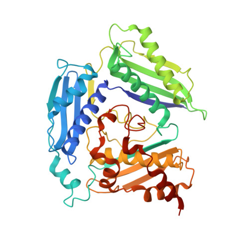
Methionine S-adenosyltransferases (MATs) are predominantly homotetramers, comprised of dimers of dimers. The larger, highly conserved intradimeric interface harbors two active sites, making the dimer the obligatory functional unit. However, functionality of the smaller, more diverged, and recently evolved interdimeric interface is largely unknown. Here, we show that the interdimeric interface of Ureaplasmaurealiticum MAT has evolved to control the catalytic activity and structural integrity of the homotetramer in response to product accumulation. When all four active sites are occupied with the product, S-adenosylmethionine (SAM), binding of four additional SAM molecules to the interdimeric interface prompts a ∼45° shift in the dimer orientation and a concomitant ∼60% increase in the interface area. This rearrangement inhibits the enzymatic activity by locking the flexible active site loops in a closed state and renders the tetramer resistant to proteolytic degradation. Our findings suggest that the interdimeric interface of MATs is subject to rapid evolutionary changes that tailor the molecular properties of the entire homotetramer to the specific needs of the organism.
- Department of Life Sciences, Ben-Gurion University of the Negev, POB 653, Beer-Sheva, 84105, Israel.
Organizational Affiliation: