


Crystal structure of CLK3 in complex with TP003
Chaikuad, A., Arrowsmith, C.H., Edwards, A.M., Bountra, C., Knapp, S., Structural Genomics Consortium (SGC)To be published.
Experimental Data Snapshot
Starting Model: experimental
View more details
Entity ID: 1 | |||||
|---|---|---|---|---|---|
| Molecule | Chains | Sequence Length | Organism | Details | Image |
| Dual specificity protein kinase CLK3 | 360 | Homo sapiens | Mutation(s): 0 Gene Names: CLK3 EC: 2.7.12.1 | 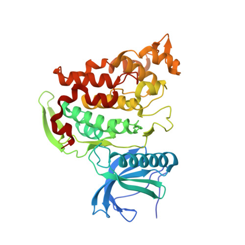 | |
UniProt & NIH Common Fund Data Resources | |||||
PHAROS: P49761 GTEx: ENSG00000179335 | |||||
Entity Groups | |||||
| Sequence Clusters | 30% Identity50% Identity70% Identity90% Identity95% Identity100% Identity | ||||
| UniProt Group | P49761 | ||||
Sequence AnnotationsExpand | |||||
Reference Sequence | |||||
| Ligands 2 Unique | |||||
|---|---|---|---|---|---|
| ID | Chains | Name / Formula / InChI Key | 2D Diagram | 3D Interactions | |
| JWN Download:Ideal Coordinates CCD File | C [auth A], J [auth B] | 4-[2-methyl-1-(4-methylpiperazin-1-yl)-1-oxidanylidene-propan-2-yl]-~{N}-(6-pyridin-4-ylimidazo[1,2-a]pyridin-2-yl)benzamide C28 H30 N6 O2 IEFFSHLHNYVSEF-UHFFFAOYSA-N |  | ||
| EDO Download:Ideal Coordinates CCD File | D [auth A] E [auth A] F [auth A] G [auth A] H [auth A] | 1,2-ETHANEDIOL C2 H6 O2 LYCAIKOWRPUZTN-UHFFFAOYSA-N |  | ||
| Length ( Å ) | Angle ( ˚ ) |
|---|---|
| a = 61.788 | α = 90 |
| b = 123.329 | β = 92.64 |
| c = 69.589 | γ = 90 |
| Software Name | Purpose |
|---|---|
| Aimless | data scaling |
| REFMAC | refinement |
| PDB_EXTRACT | data extraction |
| MOSFLM | data reduction |
| PHASER | phasing |


