6RCA

X-ray structure uridine phosphorylase from Vibrio cholerae in complex with 2.2'-anhydrouridine at 1.34 A


Experimental Data Snapshot

  • Method: X-RAY DIFFRACTION
  • Resolution: 1.34 Å
  • R-Value Free: 0.169 
  • R-Value Work: 0.135 
  • R-Value Observed: 0.136 

wwPDB Validation   3D Report Full Report


Ligand Structure Quality Assessment 


This is version 1.2 of the entry. See complete history

Re-refinement Note

This entry reflects an alternative modeling of the original data in: 5OUR


Literature

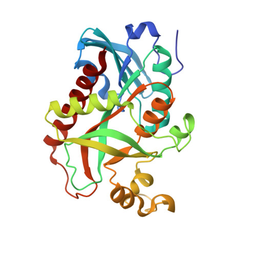
X-Ray Structure and Molecular Dynamics Study of Uridine Phosphorylase from Vibrio cholerae in Complex with 2,2'-Anhydrouridine

Eistrikh-Geller, P.A.Rubinsky, S.V.Prokofev, I.I.Gabdoulkhakov, A.G.Mironov, A.S.Lashkov, A.A.

(2020) Crystallogr Rep 


Macromolecules
Find similar proteins by:  (by identity cutoff)  |  3D Structure
Entity ID: 1
MoleculeChains Sequence LengthOrganismDetailsImage
Uridine phosphorylase
A, B, C, D, E
A, B, C, D, E, F
253Vibrio choleraeMutation(s): 0 
Gene Names: 
EC: 2.4.2.3
UniProt
Find proteins for Q9K4U1 (Vibrio cholerae)
Explore Q9K4U1 
Go to UniProtKB:  Q9K4U1
Entity Groups  
Sequence Clusters30% Identity50% Identity70% Identity90% Identity95% Identity100% Identity
UniProt GroupQ9K4U1
Sequence Annotations
Expand
  • Reference Sequence
Small Molecules
Ligands 6 Unique
IDChains Name / Formula / InChI Key2D Diagram3D Interactions
BJE
Query on BJE

Download Ideal Coordinates CCD File 
H [auth A]
I [auth B]
M [auth C]
O [auth D]
T [auth E]
H [auth A],
I [auth B],
M [auth C],
O [auth D],
T [auth E],
V [auth F]
2,2'-Anhydro-(1-beta-D-ribofuranosyl)uracil
C9 H10 N2 O5
UUGITDASWNOAGG-XVFCMESISA-N
GOL
Query on GOL

Download Ideal Coordinates CCD File 
J [auth B]
K [auth B]
P [auth D]
Q [auth D]
R [auth D]
J [auth B],
K [auth B],
P [auth D],
Q [auth D],
R [auth D],
X [auth F],
Y [auth F],
Z [auth F]
GLYCEROL
C3 H8 O3
PEDCQBHIVMGVHV-UHFFFAOYSA-N
EDO
Query on EDO

Download Ideal Coordinates CCD File 
W [auth F]1,2-ETHANEDIOL
C2 H6 O2
LYCAIKOWRPUZTN-UHFFFAOYSA-N
CL
Query on CL

Download Ideal Coordinates CCD File 
AA [auth F],
L [auth C]
CHLORIDE ION
Cl
VEXZGXHMUGYJMC-UHFFFAOYSA-M
MG
Query on MG

Download Ideal Coordinates CCD File 
U [auth E]MAGNESIUM ION
Mg
JLVVSXFLKOJNIY-UHFFFAOYSA-N
NA
Query on NA

Download Ideal Coordinates CCD File 
G [auth A],
N [auth C],
S [auth E]
SODIUM ION
Na
FKNQFGJONOIPTF-UHFFFAOYSA-N
Experimental Data & Validation

Experimental Data

  • Method: X-RAY DIFFRACTION
  • Resolution: 1.34 Å
  • R-Value Free: 0.169 
  • R-Value Work: 0.135 
  • R-Value Observed: 0.136 
  • Space Group: P 31
Unit Cell:
Length ( Å )Angle ( ˚ )
a = 93.245α = 90
b = 93.245β = 90
c = 152.928γ = 120
Software Package:
Software NamePurpose
XDSdata reduction
XSCALEdata scaling
PHASERphasing
PHENIXrefinement
PDB_EXTRACTdata extraction

Structure Validation

View Full Validation Report


Ligand Structure Quality Assessment 


Entry History & Funding Information

Deposition Data


Funding OrganizationLocationGrant Number
Other governmentRussian FederationMK-9246.2016.3.

Revision History  (Full details and data files)

  • Version 1.0: 2020-05-13
    Type: Initial release
  • Version 1.1: 2020-11-25
    Changes: Database references, Derived calculations
  • Version 1.2: 2024-01-24
    Changes: Data collection, Database references, Refinement description