6RBG

full-length bacterial polysaccharide co-polymerase


Experimental Data Snapshot

  • Method: ELECTRON MICROSCOPY
  • Resolution: 3.00 Å
  • Aggregation State: PARTICLE 
  • Reconstruction Method: SINGLE PARTICLE 

wwPDB Validation   3D Report Full Report


This is version 1.2 of the entry. See complete history


Literature

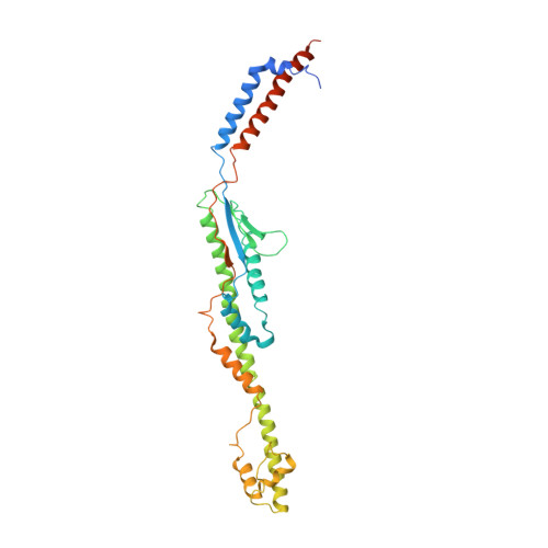
Structure of a full-length bacterial polysaccharide co-polymerase.

Wiseman, B.Nitharwal, R.G.Widmalm, G.Hogbom, M.

(2021) Nat Commun 12: 369-369

  • DOI: https://doi.org/10.1038/s41467-020-20579-1
  • Primary Citation of Related Structures:  
    6RBG

  • PubMed Abstract: 

    Lipopolysaccharides are important components of the bacterial cell envelope that among other things act as a protective barrier against the environment and toxic molecules such as antibiotics. One of the most widely disseminated pathways of polysaccharide biosynthesis is the inner membrane bound Wzy-dependent pathway. Here we present the 3.0 Å structure of the co-polymerase component of this pathway, WzzB from E. coli solved by single-particle cryo-electron microscopy. The overall architecture is octameric and resembles a box jellyfish containing a large bell-shaped periplasmic domain with the 2-helix transmembrane domain from each protomer, positioned 32 Å apart, encircling a large empty transmembrane chamber. This structure also reveals the architecture of the transmembrane domain, including the location of key residues for the Wzz-family of proteins and the Wzy-dependent pathway present in many Gram-negative bacteria, explaining several of the previous biochemical and mutational studies and lays the foundation for future investigations.


  • Organizational Affiliation

    Department of Biochemistry and Biophysics, Stockholm University, Stockholm, Sweden. bwise@dbb.su.se.


Macromolecules
Find similar proteins by:  (by identity cutoff)  |  3D Structure
Entity ID: 1
MoleculeChains Sequence LengthOrganismDetailsImage
Chain length determinant protein
A, B, C, D, E
A, B, C, D, E, F, G, H
341Escherichia coli K-12Mutation(s): 0 
Gene Names: wzzBcldrolwzzb2027JW5836
Membrane Entity: Yes 
UniProt
Find proteins for P76372 (Escherichia coli (strain K12))
Explore P76372 
Go to UniProtKB:  P76372
Entity Groups  
Sequence Clusters30% Identity50% Identity70% Identity90% Identity95% Identity100% Identity
UniProt GroupP76372
Sequence Annotations
Expand
  • Reference Sequence
Experimental Data & Validation

Experimental Data

  • Method: ELECTRON MICROSCOPY
  • Resolution: 3.00 Å
  • Aggregation State: PARTICLE 
  • Reconstruction Method: SINGLE PARTICLE 
EM Software:
TaskSoftware PackageVersion
RECONSTRUCTIONcryoSPARC2
MODEL REFINEMENTPHENIX

Structure Validation

View Full Validation Report


Entry History & Funding Information

Deposition Data


Funding OrganizationLocationGrant Number
Knut and Alice Wallenberg FoundationSweden--
Swedish Research CouncilSweden--

Revision History  (Full details and data files)

  • Version 1.0: 2020-11-18
    Type: Initial release
  • Version 1.1: 2020-11-25
    Changes: Structure summary
  • Version 1.2: 2021-01-27
    Changes: Database references