Spiro-containing derivatives show antiparasitic activity against Trypanosoma brucei through inhibition of the trypanothione reductase enzyme.
Turcano, L., Battista, T., De Haro, E.T., Missineo, A., Alli, C., Paonessa, G., Colotti, G., Harper, S., Fiorillo, A., Ilari, A., Bresciani, A.(2020) PLoS Negl Trop Dis 14: e0008339-e0008339
- PubMed: 32437349 Search on PubMedSearch on PubMed Central
- DOI: https://doi.org/10.1371/journal.pntd.0008339
- Primary Citation Related Structures:
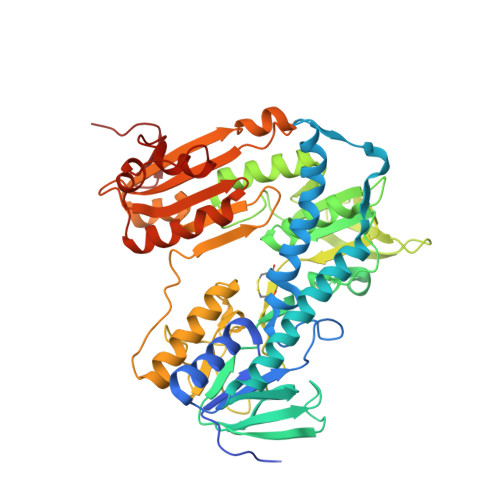
6RB5 - PubMed Abstract:
Trypanothione reductase (TR) is a key enzyme that catalyzes the reduction of trypanothione, an antioxidant dithiol that protects Trypanosomatid parasites from oxidative stress induced by mammalian host defense systems. TR is considered an attractive target for the development of novel anti-parasitic agents as it is essential for parasite survival but has no close homologue in humans. We report here the identification of spiro-containing derivatives as inhibitors of TR from Trypanosoma brucei (TbTR), the parasite responsible for Human African Trypanosomiasis. The hit series, identified by high throughput screening, was shown to bind TbTR reversibly and to compete with the trypanothione (TS2) substrate. The prototype compound 1 from this series was also found to impede the growth of Trypanosoma brucei parasites in vitro. The X-ray crystal structure of TbTR in complex with compound 1 solved at 1.98 Å allowed the identification of the hydrophobic pocket where the inhibitor binds, placed close to the catalytic histidine (His 461') and lined by Trp21, Val53, Ile106, Tyr110 and Met113. This new inhibitor is specific for TbTR and no activity was detected against the structurally similar human glutathione reductase (hGR). The central spiro scaffold is known to be suitable for brain active compounds in humans thus representing an attractive starting point for the future treatment of the central nervous system stage of T. brucei infections.
- Department of Translational and Discovery Research, Pomezia (Roma) Italy.
Organizational Affiliation: