Lack of orientation selectivity of the heme insertion in murine neuroglobin revealed by resonance Raman spectroscopy.
Milazzo, L., Exertier, C., Becucci, M., Freda, I., Montemiglio, L.C., Savino, C., Vallone, B., Smulevich, G.(2020) FEBS J 287: 4082-4097
- PubMed: 32034988 Search on PubMed
- DOI: https://doi.org/10.1111/febs.15241
- Primary Citation Related Structures:
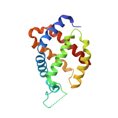
6RA6 - PubMed Abstract:
Different murine neuroglobin variants showing structural and dynamic alterations that are associated with perturbation of ligand binding have been studied: the CD loop mutants characterized by an enhanced flexibility (Gly-loop 40-48 and Gly-loop 44-47 ), the F106A mutant, and the double Gly-loop 44-47 /F106A mutant. Their ferric resonance Raman spectra in solution and in crystals are almost identical. In the high-frequency region, the identification of a double set of core size marker bands indicates the presence of two 6-coordinate low spin species. The resonance Raman data, together with the corresponding crystal structures, indicate the presence of two neuroglobin conformers with a reversed (A conformer) or a canonical (B conformer) heme insertion orientation. With the identification of the marker bands corresponding to each conformer, the data indicate that the B conformer increases at the expense of the A form, predominantly in the Gly-loop 44-47 /F106A double mutant, as confirmed by X-ray crystallography. This is the first time that a reversed heme insertion has been identified by resonance Raman in a native 6-coordinate low-spin heme protein. This diagnostic tool could be extended to other heme proteins in order to detect heme orientational disorder, which are likely to be correlated to functionally relevant heme dynamics. DATABASE: Crystallographic structure: structural data are deposited in the Protein Data Bank under the 6RA6 PDB entry.
- Dipartimento di Chimica "Ugo Schiff", Università di Firenze, Florence, Italy.
Organizational Affiliation: