6R9W | pdb_00006r9w

Crystal structure of InhA in complex with AP-124 inhibitor


Experimental Data Snapshot

  • Method: X-RAY DIFFRACTION
  • Resolution: 1.75 Å
  • R-Value Free: 
    0.227 (Depositor), 0.232 (DCC) 
  • R-Value Work: 
    0.187 (Depositor), 0.194 (DCC) 
  • R-Value Observed: 
    0.189 (Depositor) 

Starting Model: experimental
View more details

wwPDB Validation 3D Report Full Report

Validation slider image for 6R9W

Ligand Structure Quality Assessment 


This is version 1.2 of the entry. See complete history

Literature

Discovery of New and Potent InhA Inhibitors as Antituberculosis Agents: Structure-Based Virtual Screening Validated by Biological Assays and X-ray Crystallography.

Kamsri, P.Hanwarinroj, C.Phusi, N.Pornprom, T.Chayajarus, K.Punkvang, A.Suttipanta, N.Srimanote, P.Suttisintong, K.Songsiriritthigul, C.Saparpakorn, P.Hannongbua, S.Rattanabunyong, S.Seetaha, S.Choowongkomon, K.Sureram, S.Kittakoop, P.Hongmanee, P.Santanirand, P.Chen, Z.Zhu, W.Blood, R.A.Takebayashi, Y.Hinchliffe, P.Mulholland, A.J.Spencer, J.Pungpo, P.

(2020) J Chem Inf Model 60: 226-234

  • DOI: https://doi.org/10.1021/acs.jcim.9b00918
  • Primary Citation Related Structures: 
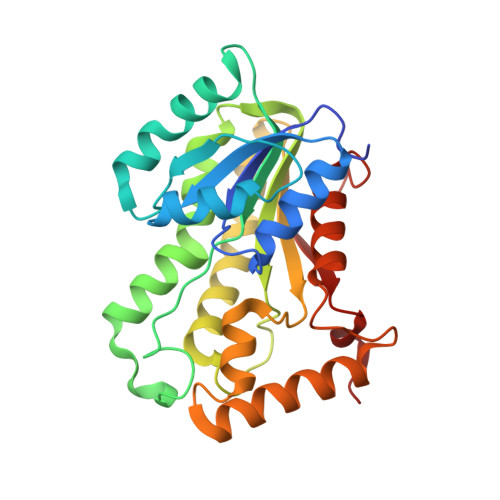
    6R9W

  • PubMed Abstract: 

    The enoyl-acyl carrier protein reductase InhA of Mycobacterium tuberculosis is an attractive, validated target for antituberculosis drug development. Moreover, direct inhibitors of InhA remain effective against InhA variants with mutations associated with isoniazid resistance, offering the potential for activity against MDR isolates. Here, structure-based virtual screening supported by biological assays was applied to identify novel InhA inhibitors as potential antituberculosis agents. High-speed Glide SP docking was initially performed against two conformations of InhA differing in the orientation of the active site Tyr158. The resulting hits were filtered for drug-likeness based on Lipinski's rule and avoidance of PAINS-like properties and finally subjected to Glide XP docking to improve accuracy. Sixteen compounds were identified and selected for in vitro biological assays, of which two (compounds 1 and 7 ) showed MIC of 12.5 and 25 μg/mL against M. tuberculosis H37Rv, respectively. Inhibition assays against purified recombinant InhA determined IC 50 values for these compounds of 0.38 and 0.22 μM, respectively. A crystal structure of the most potent compound, compound 7 , bound to InhA revealed the inhibitor to occupy a hydrophobic pocket implicated in binding the aliphatic portions of InhA substrates but distant from the NADH cofactor, i.e., in a site distinct from those occupied by the great majority of known InhA inhibitors. This compound provides an attractive starting template for ligand optimization aimed at discovery of new and effective compounds against M. tuberculosis that act by targeting InhA.


  • Organizational Affiliation
    • Division of Chemistry, Faculty of Science , Nakhon Phanom University , 48000 Nakhon Phanom , Thailand.

Macromolecule Content 

  • Total Structure Weight: 177.31 kDa 
  • Atom Count: 13,090 
  • Modeled Residue Count: 1,571 
  • Deposited Residue Count: 1,632 
  • Unique protein chains: 1

Macromolecules

Find similar proteins by:|  3D Structure
Entity ID: 1
MoleculeChains  Sequence LengthOrganismDetailsImage
Enoyl-[acyl-carrier-protein] reductase [NADH]
A, B, C, D, E
A, B, C, D, E, F
272Mycobacterium tuberculosis H37RvMutation(s): 0 
Gene Names: inhARv1484MTCY277.05
EC: 1.3.1.9
UniProt
Find proteins for P9WGR1 (Mycobacterium tuberculosis (strain ATCC 25618 / H37Rv))
Explore P9WGR1 
Go to UniProtKB:  P9WGR1
Entity Groups
Sequence Clusters30% Identity50% Identity70% Identity90% Identity95% Identity100% Identity
UniProt GroupP9WGR1
Sequence Annotations
Expand
Reference Sequence

Small Molecules

Ligands 2 Unique
IDChains Name / Formula / InChI Key2D Diagram3D Interactions
NAD

Query on NAD


Download:Ideal Coordinates CCD File
H [auth A]
I [auth B]
J [auth C]
K [auth D]
L [auth E]
H [auth A],
I [auth B],
J [auth C],
K [auth D],
L [auth E],
M [auth F]
NICOTINAMIDE-ADENINE-DINUCLEOTIDE
C21 H27 N7 O14 P2
BAWFJGJZGIEFAR-NNYOXOHSSA-N
JVZ
(Subject of Investigation/LOI)

Query on JVZ


Download:Ideal Coordinates CCD File
G [auth A](2~{S})-1-(benzimidazol-1-yl)-3-(2,3-dihydro-1~{H}-inden-5-yloxy)propan-2-ol
C19 H20 N2 O2
OZCCXUVVMPVMIB-INIZCTEOSA-N

Experimental Data & Validation

Experimental Data

  • Method: X-RAY DIFFRACTION
  • Resolution: 1.75 Å
  • R-Value Free:  0.227 (Depositor), 0.232 (DCC) 
  • R-Value Work:  0.187 (Depositor), 0.194 (DCC) 
  • R-Value Observed: 0.189 (Depositor) 
Space Group: C 1 2 1
Unit Cell:
Length ( Å )Angle ( ˚ )
a = 100.9α = 90
b = 81.602β = 95.52
c = 189.415γ = 90
Software Package:
Software NamePurpose
PHENIXrefinement
XDSdata reduction
Aimlessdata scaling
PHASERphasing

Structure Validation

View Full Validation Report


Ligand Structure Quality Assessment 


Entry History 

& Funding Information

Deposition Data


Funding OrganizationLocationGrant Number
Engineering and Physical Sciences Research CouncilUnited KingdomEP/M027546/1
Biotechnology and Biological Sciences Research CouncilUnited KingdomBB/L01386X/1
Biotechnology and Biological Sciences Research CouncilUnited KingdomBB/M012107/1

Revision History  (Full details and data files)

  • Version 1.0: 2019-12-25
    Type: Initial release
  • Version 1.1: 2020-02-05
    Changes: Database references
  • Version 1.2: 2024-01-24
    Changes: Data collection, Database references, Refinement description