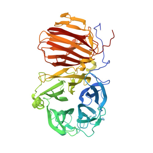
6R3U

Endo-levanase BT1760 mutant E221A from Bacteroides thetaiotaomicron complexed with levantetraose


Experimental Data Snapshot

  • Method: X-RAY DIFFRACTION
  • Resolution: 1.90 Å
  • R-Value Free: 0.190 
  • R-Value Work: 0.161 

wwPDB Validation   3D Report Full Report


This is version 2.1 of the entry. See complete history


Literature

First crystal structure of an endo-levanase - the BT1760 from a human gut commensal Bacteroides thetaiotaomicron.

Ernits, K.Eek, P.Lukk, T.Visnapuu, T.Alamae, T.

(2019) Sci Rep 9: 8443-8443

  • DOI: https://doi.org/10.1038/s41598-019-44785-0
  • Primary Citation of Related Structures:  
    6R3R, 6R3U

  • PubMed Abstract: 

    The endo-levanase BT1760 of a human gut commensal Bacteroides thetaiotaomicron randomly cuts a β-2,6-linked fructan, levan, into fructo-oligosaccharides providing a prebiotic substrate for gut microbiota. Here we introduce the crystal structure of BT1760 at resolution of 1.65 Å. The fold of the enzyme is typical for GH32 family proteins: a catalytic N-terminal five-bladed β-propeller connected with a C-terminal β-sandwich domain. The levantetraose-bound structure of catalytically inactive mutant E221A at 1.90-Å resolution reveals differences in substrate binding between the endo-acting fructanases. A shallow substrate-binding pocket of the endo-inulinase INU2 of Aspergillus ficuum binds at least three fructose residues at its flat bottom. In the levantetraose-soaked crystal of the endo-levanase E221A mutant the ligand was bent into the pond-like substrate pocket with its fructose residues making contacts at -3, -2, -1 and + 1 subsites residing at several pocket depths. Binding of levantetraose to the β-sandwich domain was not detected. The N- and C-terminal modules of BT1760 did not bind levan if expressed separately, the catalytic domain lost its activity and both modules tended to precipitate. We gather that endo-levanase BT1760 requires both domains for correct folding, solubility and stability of the protein.


  • Organizational Affiliation

    Department of Genetics, Institute of Molecular and Cell Biology, University of Tartu, Riia 23, 51010, Tartu, Estonia.


Macromolecules
Find similar proteins by:  (by identity cutoff)  |  3D Structure
Entity ID: 1
MoleculeChains Sequence LengthOrganismDetailsImage
Glycoside hydrolase family 32508Bacteroides thetaiotaomicronMutation(s): 1 
Gene Names: BT_1760
UniProt
Find proteins for Q8A6W6 (Bacteroides thetaiotaomicron (strain ATCC 29148 / DSM 2079 / JCM 5827 / CCUG 10774 / NCTC 10582 / VPI-5482 / E50))
Explore Q8A6W6 
Go to UniProtKB:  Q8A6W6
Entity Groups  
Sequence Clusters30% Identity50% Identity70% Identity90% Identity95% Identity100% Identity
UniProt GroupQ8A6W6
Sequence Annotations
Expand
  • Reference Sequence
Oligosaccharides

Help

Entity ID: 2
MoleculeChains Length2D Diagram Glycosylation3D Interactions
beta-D-fructofuranose-(2-6)-beta-D-fructofuranose-(2-6)-beta-D-fructofuranose-(2-6)-beta-D-fructofuranose
B
4N/A
Glycosylation Resources
GlyTouCan:  G94951WG
GlyCosmos:  G94951WG
Experimental Data & Validation

Experimental Data

  • Method: X-RAY DIFFRACTION
  • Resolution: 1.90 Å
  • R-Value Free: 0.190 
  • R-Value Work: 0.161 
  • Space Group: I 2 2 2
Unit Cell:
Length ( Å )Angle ( ˚ )
a = 78.114α = 90
b = 112.334β = 90
c = 174.135γ = 90
Software Package:
Software NamePurpose
XDSdata reduction
PHENIXrefinement
XDSdata scaling

Structure Validation

View Full Validation Report


Entry History & Funding Information

Deposition Data


Funding OrganizationLocationGrant Number
Estonian Research CouncilEstoniaPUT1050
Estonian Research CouncilEstoniaIUT19-9

Revision History  (Full details and data files)

  • Version 1.0: 2019-06-19
    Type: Initial release
  • Version 1.1: 2019-06-26
    Changes: Data collection, Database references
  • Version 2.0: 2020-07-29
    Type: Remediation
    Reason: Carbohydrate remediation
    Changes: Atomic model, Data collection, Derived calculations, Structure summary
  • Version 2.1: 2024-01-24
    Changes: Data collection, Database references, Derived calculations, Refinement description, Structure summary